ARG63469
anti-HEC1 antibody
anti-HEC1 antibody for IHC-Formalin-fixed paraffin-embedded sections,Western blot and Human
Cancer antibody; Cell Biology and Cellular Response antibody; Gene Regulation antibody
Overview
| Product Description | Goat Polyclonal antibody recognizes HEC1 |
|---|---|
| Tested Reactivity | Hu |
| Tested Application | IHC-P, WB |
| Host | Goat |
| Clonality | Polyclonal |
| Isotype | IgG |
| Target Name | HEC1 |
| Antigen Species | Human |
| Immunogen | C-YEKKATLIKSSEE |
| Conjugation | Un-conjugated |
| Alternate Names | Kinetochore-associated protein 2; TID3; hsNDC80; HEC1; KNTC2; Kinetochore protein NDC80 homolog; Highly expressed in cancer protein; HEC; Retinoblastoma-associated protein HEC; HsHec1; Kinetochore protein Hec1 |
Application Instructions
| Application Suggestion |
|
||||||
|---|---|---|---|---|---|---|---|
| Application Note | WB: Recommend incubate at RT for 1h. IHC-P: Antigen Retrieval: Steam tissue section in Citrate buffer (pH 6.0). * The dilutions indicate recommended starting dilutions and the optimal dilutions or concentrations should be determined by the scientist. |
Properties
| Form | Liquid |
|---|---|
| Purification | Purified from goat serum by antigen affinity chromatography. |
| Buffer | Tris saline (pH 7.3), 0.02% Sodium azide and 0.5% BSA. |
| Preservative | 0.02% Sodium azide |
| Stabilizer | 0.5% BSA |
| Concentration | 0.5 mg/ml |
| Storage Instruction | For continuous use, store undiluted antibody at 2-8°C for up to a week. For long-term storage, aliquot and store at -20°C or below. Storage in frost free freezers is not recommended. Avoid repeated freeze/thaw cycles. Suggest spin the vial prior to opening. The antibody solution should be gently mixed before use. |
| Note | For laboratory research only, not for drug, diagnostic or other use. |
Bioinformation
| Database Links | |
|---|---|
| Background | This gene encodes a component of the NDC80 kinetochore complex. The encoded protein consists of an N-terminal microtubule binding domain and a C-terminal coiled-coiled domain that interacts with other components of the complex. This protein functions to organize and stabilize microtubule-kinetochore interactions and is required for proper chromosome segregation. [provided by RefSeq, Oct 2011] |
| Research Area | Cancer antibody; Cell Biology and Cellular Response antibody; Gene Regulation antibody |
| Calculated MW | 74 kDa |
| PTM | Phosphorylation begins in S phase of the cell cycle and peaks in mitosis. Phosphorylated by NEK2. May also be phosphorylated by AURKA and AURKB. |
Images (3) Click the Picture to Zoom In
-
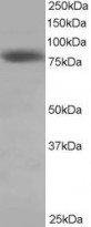
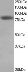
ARG63469 anti-HEC1 antibody WB image
Western Blot: HeLa lysate (RIPA buffer, 35 µg total protein per lane) stained with ARG63469 anti-HEC1 antibody at 0.5 µg/ml dilution.
-
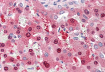
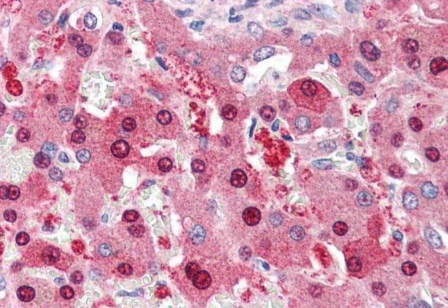
ARG63469 anti-HEC1 antibody IHC-P image
Immunohistochemistry: Paraffin-embedded Human liver tissue. Antigen Retrieval: Steam tissue section in Citrate buffer (pH 6.0). The tissue section was stained with ARG63469 anti-HEC1 antibody at 5 µg/ml dilution followed by AP-staining.
-
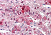
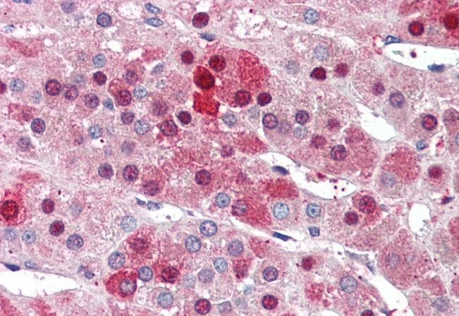
ARG63469 anti-HEC1 antibody IHC-P image
Immunohistochemistry: Paraffin-embedded Human adrenal gland tissue. Antigen Retrieval: Steam tissue section in Citrate buffer (pH 6.0). The tissue section was stained with ARG63469 anti-HEC1 antibody at 5 µg/ml dilution followed by AP-staining.