ARG66174
anti-EFHD1 antibody
anti-EFHD1 antibody for ICC/IF,IHC-Formalin-fixed paraffin-embedded sections and Human,Mouse,Rat
Overview
| Product Description | Mouse Monoclonal antibody recognizes EFHD1 |
|---|---|
| Tested Reactivity | Hu, Ms, Rat |
| Tested Application | ICC/IF, IHC-P |
| Specificity | The antibody detects endogenous EFHD1 proteins. |
| Host | Mouse |
| Clonality | Monoclonal |
| Target Name | EFHD1 |
| Antigen Species | Human |
| Immunogen | Synthetic peptide of Human EFHD1. |
| Conjugation | Un-conjugated |
| Alternate Names | EF-hand domain-containing protein D1; Swiprosin-2; EF-hand domain-containing protein 1; MSTP133; MST133; SWS2; PP3051 |
Application Instructions
| Application Suggestion |
|
||||||
|---|---|---|---|---|---|---|---|
| Application Note | IHC-P: Antigen Retrieval: Boil tissue section in Sodium citrate buffer (pH 6.0) for 20 min. * The dilutions indicate recommended starting dilutions and the optimal dilutions or concentrations should be determined by the scientist. |
Properties
| Form | Liquid |
|---|---|
| Purification | Affinity purification with immunogen. |
| Buffer | PBS (pH 7.4), 0.02% Sodium azide and 50% Glycerol. |
| Preservative | 0.02% Sodium azide |
| Stabilizer | 50% Glycerol |
| Concentration | 1 mg/ml |
| Storage Instruction | For continuous use, store undiluted antibody at 2-8°C for up to a week. For long-term storage, aliquot and store at -20°C. Storage in frost free freezers is not recommended. Avoid repeated freeze/thaw cycles. Suggest spin the vial prior to opening. The antibody solution should be gently mixed before use. |
| Note | For laboratory research only, not for drug, diagnostic or other use. |
Bioinformation
| Database Links |
Swiss-port # Q9BUP0 Human EF-hand domain-containing protein D1 Swiss-port # Q9D4J1 Mouse EF-hand domain-containing protein D1 |
|---|---|
| Gene Symbol | EFHD1 |
| Gene Full Name | EF-hand domain family, member D1 |
| Background | EFHD1 is an EF-hand domain-containing protein that displays increased expression during neuronal differentiation (Tominaga and Tomooka, 2002 [PubMed 12270117]).[supplied by OMIM, Mar 2008] |
| Calculated MW | 27 kDa |
Images (22) Click the Picture to Zoom In
-

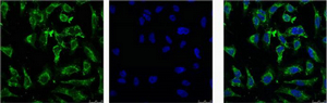
ARG66174 anti-EFHD1 antibody ICC/IF image
Immunofluorescence: HeLa cells stained with ARG66174 anti-EFHD1 antibody (green).
Left: Target. Middle: DAPI. Right: merge image.
-
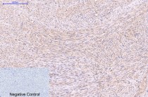
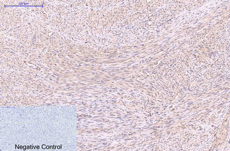
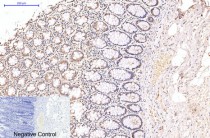
ARG66174 anti-EFHD1 antibody IHC-P image
Immunohistochemistry: Paraffin-embedded Human uterus tissue stained with ARG66174 anti-EFHD1 antibody at 1:200 dilution (4°C, overnight). Antigen Retrieval: Boil tissue section in Sodium citrate buffer (pH 6.0) for 20 min.
Negative control was used by secondary antibody only.
-
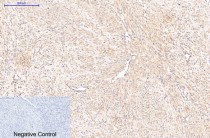
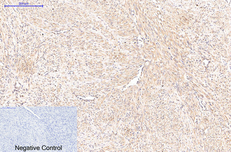
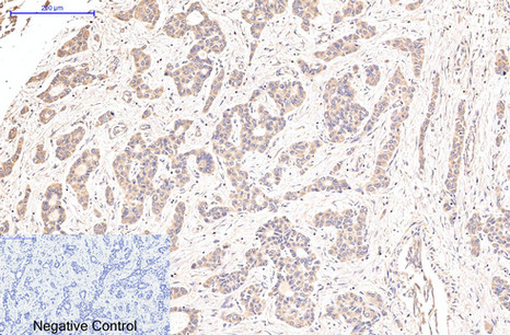
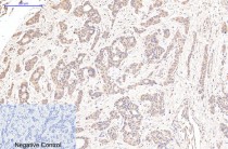
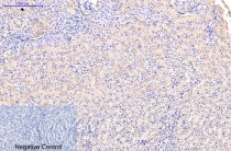
ARG66174 anti-EFHD1 antibody IHC-P image
Immunohistochemistry: Paraffin-embedded Human uterus cancer tissue stained with ARG66174 anti-EFHD1 antibody at 1:200 dilution (4°C, overnight). Antigen Retrieval: Boil tissue section in Sodium citrate buffer (pH 6.0) for 20 min.
Negative control was used by secondary antibody only.
-
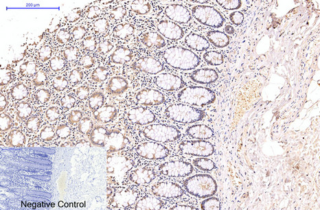
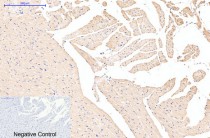
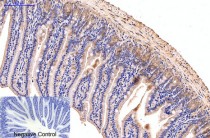
ARG66174 anti-EFHD1 antibody IHC-P image
Immunohistochemistry: Paraffin-embedded Human colon tissue stained with ARG66174 anti-EFHD1 antibody at 1:200 dilution (4°C, overnight). Antigen Retrieval: Boil tissue section in Sodium citrate buffer (pH 6.0) for 20 min.
Negative control was used by secondary antibody only.
-
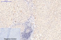
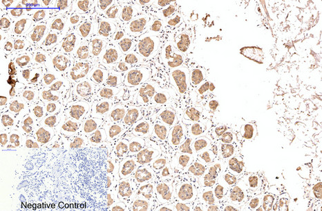
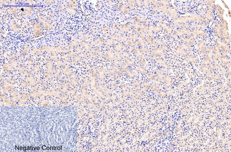
ARG66174 anti-EFHD1 antibody IHC-P image
Immunohistochemistry: Paraffin-embedded Human liver tissue stained with ARG66174 anti-EFHD1 antibody at 1:200 dilution (4°C, overnight). Antigen Retrieval: Boil tissue section in Sodium citrate buffer (pH 6.0) for 20 min.
Negative control was used by secondary antibody only.
-
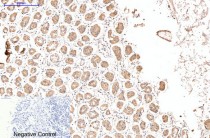
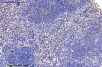
ARG66174 anti-EFHD1 antibody IHC-P image
Immunohistochemistry: Paraffin-embedded Human liver cancer tissue stained with ARG66174 anti-EFHD1 antibody at 1:200 dilution (4°C, overnight). Antigen Retrieval: Boil tissue section in Sodium citrate buffer (pH 6.0) for 20 min.
Negative control was used by secondary antibody only.
-
ARG66174 anti-EFHD1 antibody IHC-P image
Immunohistochemistry: Paraffin-embedded Human stomach tissue stained with ARG66174 anti-EFHD1 antibody at 1:200 dilution (4°C, overnight). Antigen Retrieval: Boil tissue section in Sodium citrate buffer (pH 6.0) for 20 min.
Negative control was used by secondary antibody only.
-
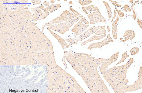
ARG66174 anti-EFHD1 antibody IHC-P image
Immunohistochemistry: Paraffin-embedded Rat heart tissue stained with ARG66174 anti-EFHD1 antibody at 1:200 dilution (4°C, overnight). Antigen Retrieval: Boil tissue section in Sodium citrate buffer (pH 6.0) for 20 min.
Negative control was used by secondary antibody only.
-
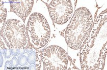
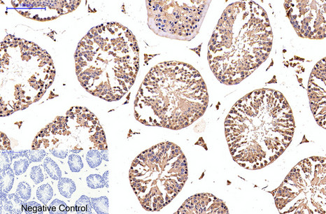
ARG66174 anti-EFHD1 antibody IHC-P image
Immunohistochemistry: Paraffin-embedded Rat testis tissue stained with ARG66174 anti-EFHD1 antibody at 1:200 dilution (4°C, overnight). Antigen Retrieval: Boil tissue section in Sodium citrate buffer (pH 6.0) for 20 min.
Negative control was used by secondary antibody only.
-

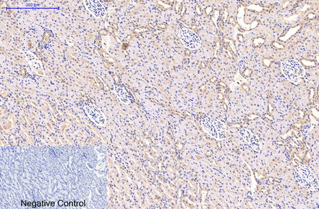
ARG66174 anti-EFHD1 antibody IHC-P image
Immunohistochemistry: Paraffin-embedded Rat kidney tissue stained with ARG66174 anti-EFHD1 antibody at 1:200 dilution (4°C, overnight). Antigen Retrieval: Boil tissue section in Sodium citrate buffer (pH 6.0) for 20 min.
Negative control was used by secondary antibody only.
-
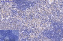
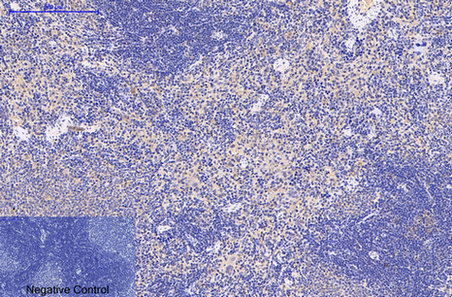
ARG66174 anti-EFHD1 antibody IHC-P image
Immunohistochemistry: Paraffin-embedded Rat spleen tissue stained with ARG66174 anti-EFHD1 antibody at 1:200 dilution (4°C, overnight). Antigen Retrieval: Boil tissue section in Sodium citrate buffer (pH 6.0) for 20 min.
Negative control was used by secondary antibody only.
-
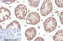
ARG66174 anti-EFHD1 antibody IHC-P image
Immunohistochemistry: Paraffin-embedded Mouse testis tissue stained with ARG66174 anti-EFHD1 antibody at 1:200 dilution (4°C, overnight). Antigen Retrieval: Boil tissue section in Sodium citrate buffer (pH 6.0) for 20 min.
Negative control was used by secondary antibody only.
-
ARG66174 anti-EFHD1 antibody IHC-P image
Immunohistochemistry: Paraffin-embedded Mouse colon tissue stained with ARG66174 anti-EFHD1 antibody at 1:200 dilution (4°C, overnight). Antigen Retrieval: Boil tissue section in Sodium citrate buffer (pH 6.0) for 20 min.
Negative control was used by secondary antibody only.
-
ARG66174 anti-EFHD1 antibody IHC-P image
Immunohistochemistry: Paraffin-embedded Mouse kidney tissue stained with ARG66174 anti-EFHD1 antibody at 1:200 dilution (4°C, overnight). Antigen Retrieval: Boil tissue section in Sodium citrate buffer (pH 6.0) for 20 min.
Negative control was used by secondary antibody only.
-
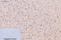
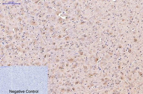
ARG66174 anti-EFHD1 antibody IHC-P image
Immunohistochemistry: Paraffin-embedded Mouse brain tissue stained with ARG66174 anti-EFHD1 antibody at 1:200 dilution (4°C, overnight). Antigen Retrieval: Boil tissue section in Sodium citrate buffer (pH 6.0) for 20 min.
Negative control was used by secondary antibody only.
-
ARG66174 anti-EFHD1 antibody IHC-P image
Immunohistochemistry: Paraffin-embedded Mouse spleen tissue stained with ARG66174 anti-EFHD1 antibody at 1:200 dilution (4°C, overnight). Antigen Retrieval: Boil tissue section in Sodium citrate buffer (pH 6.0) for 20 min.
Negative control was used by secondary antibody only.
-
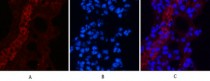
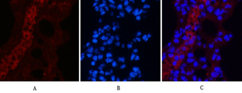
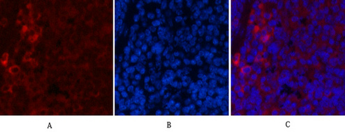
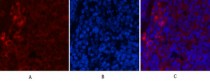
ARG66174 anti-EFHD1 antibody IHC image
Immunohistochemistry: Mouse lung tissue stained with ARG66174 anti-EFHD1 antibody (red) at 1:200 dilution (4°C, overnight).
Picture A: Target. Picture B: DAPI. Picture C: merge of A+B.
-
ARG66174 anti-EFHD1 antibody IHC image
Immunohistochemistry: Mouse lung tissue stained with ARG66174 anti-EFHD1 antibody (red) at 1:200 dilution (4°C, overnight).
Picture A: Target. Picture B: DAPI. Picture C: merge of A+B.
-
ARG66174 anti-EFHD1 antibody IHC image
Immunohistochemistry: Mouse lung tissue stained with ARG66174 anti-EFHD1 antibody (red) at 1:200 dilution (4°C, overnight).
Picture A: Target. Picture B: DAPI. Picture C: merge of A+B.
-
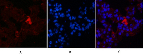
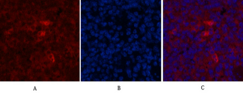
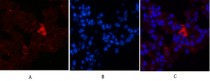
ARG66174 anti-EFHD1 antibody IHC image
Immunohistochemistry: Mouse spleen tissue stained with ARG66174 anti-EFHD1 antibody (red) at 1:200 dilution (4°C, overnight).
Picture A: Target. Picture B: DAPI. Picture C: merge of A+B.
-
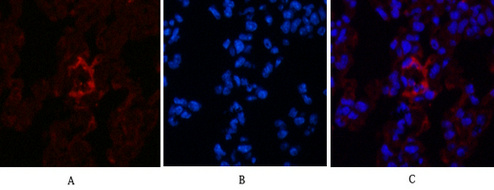
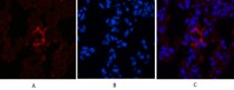
ARG66174 anti-EFHD1 antibody IHC image
Immunohistochemistry: Mouse spleen tissue stained with ARG66174 anti-EFHD1 antibody (red) at 1:200 dilution (4°C, overnight).
Picture A: Target. Picture B: DAPI. Picture C: merge of A+B.
-

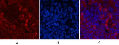
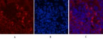
ARG66174 anti-EFHD1 antibody IHC image
Immunohistochemistry: Mouse spleen tissue stained with ARG66174 anti-EFHD1 antibody (red) at 1:200 dilution (4°C, overnight).
Picture A: Target. Picture B: DAPI. Picture C: merge of A+B.