ARG66662
anti-Caspase 8 antibody
anti-Caspase 8 antibody for IHC-Formalin-fixed paraffin-embedded sections,Western blot and Human,Mouse,Rat
Overview
| Product Description | Mouse Monoclonal antibody recognizes Caspase 8 |
|---|---|
| Tested Reactivity | Hu, Ms, Rat |
| Tested Application | IHC-P, WB |
| Host | Mouse |
| Clonality | Monoclonal |
| Isotype | IgG |
| Target Name | Caspase 8 |
| Antigen Species | Human |
| Immunogen | Recombinant protein of Human Caspase 8. |
| Conjugation | Un-conjugated |
| Alternate Names | Casp-8; FADD-like ICE; EC 3.4.22.61; CAP4; ICE-like apoptotic protease 5; MORT1-associated ced-3 homolog; FLICE; Apoptotic cysteine protease; FADD-homologous ICE/ced-3-like protease; Caspase-8; Apoptotic protease Mch-5; CASP-8; MCH5; ALPS2B; MACH |
Application Instructions
| Application Suggestion |
|
||||||
|---|---|---|---|---|---|---|---|
| Application Note | IHC-P: Antigen Retrieval: Boil tissue section in Sodium citrate buffer (pH 6.0) for 20 min. * The dilutions indicate recommended starting dilutions and the optimal dilutions or concentrations should be determined by the scientist. |
||||||
| Observed Size | ~ 55 kDa |
Properties
| Form | Liquid |
|---|---|
| Purification | Affinity purification with immunogen. |
| Buffer | PBS (pH 7.4), 0.02% Sodium azide and 50% Glycerol. |
| Preservative | 0.02% Sodium azide |
| Stabilizer | 50% Glycerol |
| Concentration | 1 mg/ml |
| Storage Instruction | For continuous use, store undiluted antibody at 2-8°C for up to a week. For long-term storage, aliquot and store at -20°C. Storage in frost free freezers is not recommended. Avoid repeated freeze/thaw cycles. Suggest spin the vial prior to opening. The antibody solution should be gently mixed before use. |
| Note | For laboratory research only, not for drug, diagnostic or other use. |
Bioinformation
| Database Links | |
|---|---|
| Gene Symbol | CASP8 |
| Gene Full Name | caspase 8, apoptosis-related cysteine peptidase |
| Background | This gene encodes a member of the cysteine-aspartic acid protease (caspase) family. Sequential activation of caspases plays a central role in the execution-phase of cell apoptosis. Caspases exist as inactive proenzymes composed of a prodomain, a large protease subunit, and a small protease subunit. Activation of caspases requires proteolytic processing at conserved internal aspartic residues to generate a heterodimeric enzyme consisting of the large and small subunits. This protein is involved in the programmed cell death induced by Fas and various apoptotic stimuli. The N-terminal FADD-like death effector domain of this protein suggests that it may interact with Fas-interacting protein FADD. This protein was detected in the insoluble fraction of the affected brain region from Huntington disease patients but not in those from normal controls, which implicated the role in neurodegenerative diseases. Many alternatively spliced transcript variants encoding different isoforms have been described, although not all variants have had their full-length sequences determined. [provided by RefSeq, Jul 2008] |
| Function | Most upstream protease of the activation cascade of caspases responsible for the TNFRSF6/FAS mediated and TNFRSF1A induced cell death. Binding to the adapter molecule FADD recruits it to either receptor. The resulting aggregate called death-inducing signaling complex (DISC) performs CASP8 proteolytic activation. The active dimeric enzyme is then liberated from the DISC and free to activate downstream apoptotic proteases. Proteolytic fragments of the N-terminal propeptide (termed CAP3, CAP5 and CAP6) are likely retained in the DISC. Cleaves and activates CASP3, CASP4, CASP6, CASP7, CASP9 and CASP10. May participate in the GZMB apoptotic pathways. Cleaves ADPRT. Hydrolyzes the small-molecule substrate, Ac-Asp-Glu-Val-Asp-|-AMC. Likely target for the cowpox virus CRMA death inhibitory protein. Isoform 5, isoform 6, isoform 7 and isoform 8 lack the catalytic site and may interfere with the pro-apoptotic activity of the complex. [UniProt] |
| Cellular Localization | Cytoplasm. [UniProt] |
| Highlight | Related news: Ripoptosome & Necrosome antibody panels are launched |
| Calculated MW | 55 kDa |
| PTM | Generation of the subunits requires association with the death-inducing signaling complex (DISC), whereas additional processing is likely due to the autocatalytic activity of the activated protease. GZMB and CASP10 can be involved in these processing events. Phosphorylation on Ser-387 during mitosis by CDK1 inhibits activation by proteolysis and prevents apoptosis. This phosphorylation occurs in cancer cell lines, as well as in primary breast tissues and lymphocytes. [UniProt] |
Images (5) Click the Picture to Zoom In
-
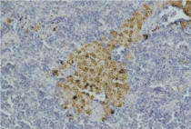
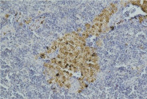
ARG66662 anti-Caspase 8 antibody IHC-P image
Immunohistochemistry: Paraffin-embedded Mouse spleen tissue stained with ARG66662 anti-Caspase 8 antibody.
-

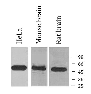
ARG66662 anti-Caspase 8 antibody WB image
Western blot: HeLa, Mouse brain and Rat brain lysates stained with ARG66662 anti-Caspase 8 antibody.
-
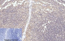
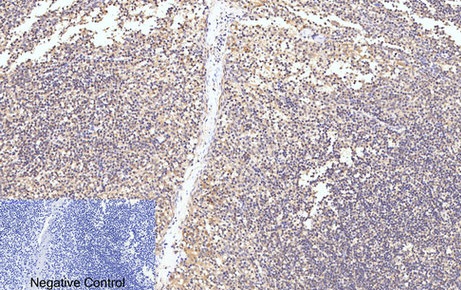
ARG66662 anti-Caspase 8 antibody IHC-P image
Immunohistochemistry: Paraffin-embedded Human tonsil tissue stained with ARG66662 anti-Caspase 8 antibody at 1:200 dilution, overnight at 4°C. Antigen Retrieval: Boil tissue section in Sodium citrate buffer (pH 6.0) for 20 min. Negative control was used by secondary antibody only.
-
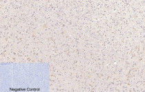
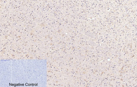
ARG66662 anti-Caspase 8 antibody IHC-P image
Immunohistochemistry: Paraffin-embedded Mouse brain tissue stained with ARG66662 anti-Caspase 8 antibody at 1:200 dilution, overnight at 4°C. Antigen Retrieval: Boil tissue section in Sodium citrate buffer (pH 6.0) for 20 min. Negative control was used by secondary antibody only.
-

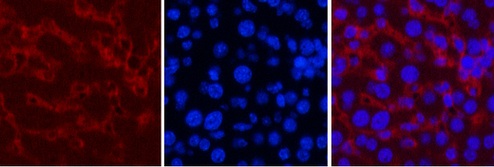
ARG66662 anti-Caspase 8 antibody IHC-P image
Immunohistochemistry: Paraffin-embedded Mouse liver tissue stained with ARG66662 anti-Caspase 8 antibody (red) at 1:200 dilution, overnight at 4°C. DAPI (blue) for nuclear staining.