ARG22899
anti-CD8 antibody [YTS105.18]
anti-CD8 antibody [YTS105.18] for Flow cytometry,ICC/IF,IHC-Frozen sections and Mouse
Developmental Biology antibody; Immune System antibody; Cytotoxic T antibody; Cytotoxic T Cell Surface Study antibody; Tumor-infiltrating Lymphocyte Study antibody
Overview
| Product Description | Rat Monoclonal antibody [YTS105.18] recognizes CD8 Rat anti Mouse CD8, clone YTS105.18 recognizes a non polymorphic epitope on the mouse CD8 alpha chain. This antibody has been reported to block MHC I dependent T cell responses in vitro and in vivo, and induces transplantation tolerance in combination with CD4 antibodies (Cobbold et al. 1990 & Wise et al. 1998). |
|---|---|
| Tested Reactivity | Ms |
| Tested Application | FACS, ICC/IF, IHC-Fr |
| Host | Rat |
| Clonality | Monoclonal |
| Clone | YTS105.18 |
| Isotype | IgG2a |
| Target Name | CD8 |
| Antigen Species | Mouse |
| Immunogen | Mouse spleen cells. |
| Conjugation | Un-conjugated |
| Alternate Names | T-cell surface glycoprotein CD8 alpha chain; Leu2; p32; T-lymphocyte differentiation antigen T8/Leu-2; CD8; MAL; CD antigen CD8a |
Application Instructions
| Application Suggestion |
|
||||||||
|---|---|---|---|---|---|---|---|---|---|
| Application Note | FACS: Use 10 µl of the suggested working dilution to label 10^6 cells in 100 µl. * The dilutions indicate recommended starting dilutions and the optimal dilutions or concentrations should be determined by the scientist. |
Properties
| Form | Liquid |
|---|---|
| Purification | Purification with Protein G. |
| Buffer | PBS and 0.09% Sodium azide |
| Preservative | 0.09% Sodium azide |
| Concentration | 1 mg/ml |
| Storage Instruction | For continuous use, store undiluted antibody at 2-8°C for up to a week. For long-term storage, aliquot and store at -20°C or below. Storage in frost free freezers is not recommended. Avoid repeated freeze/thaw cycles. Suggest spin the vial prior to opening. The antibody solution should be gently mixed before use. |
| Note | For laboratory research only, not for drug, diagnostic or other use. |
Bioinformation
| Database Links |
Swiss-port # P01731 Mouse T-cell surface glycoprotein CD8 alpha chain |
|---|---|
| Gene Symbol | Cd8a |
| Gene Full Name | CD8 antigen, alpha chain |
| Background | CD8 antigen is a cell surface glycoprotein found on most cytotoxic T lymphocytes that mediates efficient cell-cell interactions within the immune system. The CD8 antigen acts as a coreceptor with the T-cell receptor on the T lymphocyte to recognize antigens displayed by an antigen presenting cell in the context of class I MHC molecules. The coreceptor functions as either a homodimer composed of two alpha chains or as a heterodimer composed of one alpha and one beta chain. Both alpha and beta chains share significant homology to immunoglobulin variable light chains. This gene encodes the CD8 alpha chain. Multiple transcript variants encoding different isoforms have been found for this gene. [provided by RefSeq, Nov 2011] |
| Function | CD8 is an integral membrane glycoprotein that plays an essential role in the immune response and serves multiple functions in responses against both external and internal offenses. In T-cells, functions primarily as a coreceptor for MHC class I molecule:peptide complex. The antigens presented by class I peptides are derived from cytosolic proteins while class II derived from extracellular proteins. Interacts simultaneously with the T-cell receptor (TCR) and the MHC class I proteins presented by antigen presenting cells (APCs). In turn, recruits the Src kinase LCK to the vicinity of the TCR-CD3 complex. LCK then initiates different intracellular signaling pathways by phosphorylating various substrates ultimately leading to lymphokine production, motility, adhesion and activation of cytotoxic T-lymphocytes (CTLs). This mechanism enables CTLs to recognize and eliminate infected cells and tumor cells. In NK-cells, the presence of CD8A homodimers at the cell surface provides a survival mechanism allowing conjugation and lysis of multiple target cells. CD8A homodimer molecules also promote the survival and differentiation of activated lymphocytes into memory CD8 T-cells. [UniProt] |
| Highlight | Related products: CD8 antibodies; CD8 ELISA Kits; CD8 Duos / Panels; Anti-Rat IgG secondary antibodies; Related news: New antibody panels and duos for Tumor immune microenvironment Tumor-Infiltrating Lymphocytes (TILs) Detecting exosomal HMGB1 for ICD research |
| Research Area | Developmental Biology antibody; Immune System antibody; Cytotoxic T antibody; Cytotoxic T Cell Surface Study antibody; Tumor-infiltrating Lymphocyte Study antibody |
| Calculated MW | 26 kDa |
| PTM | All of the five most C-terminal cysteines form inter-chain disulfide bonds in dimers and higher multimers, while the four N-terminal cysteines do not. |
Images (10) Click the Picture to Zoom In
-
ARG22899 anti-CD8 antibody [YTS105.18] FACS image
Flow Cytometry: Mouse spleen cells stained with ARG22899 anti-CD8 antibody [YTS105.18].
-
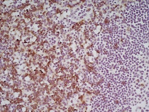
ARG22899 anti-CD8 antibody [YTS105.18] IHC-Fr image
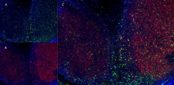
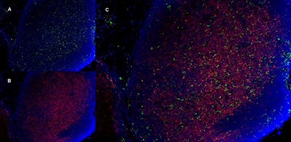
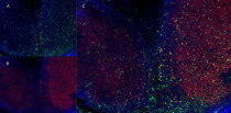
Immunohistochemistry: Mouse lymph node cryosection stained with Rat anti Mouse CD11b antibody, green in A and ARG22899 anti-CD8 antibody [YTS105.18], red in B, Merged image in C.
-
ARG22899 anti-CD8 antibody [YTS105.18] IHC-Fr image
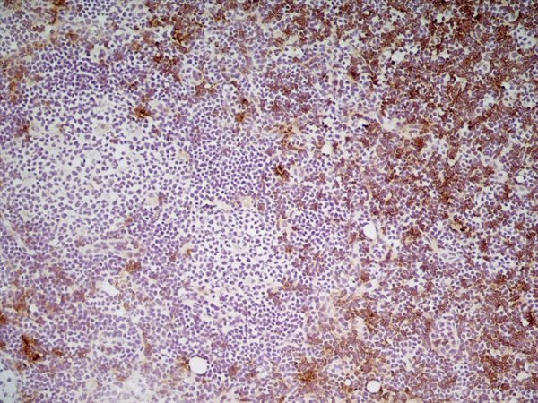
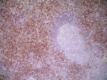
Immunohistochemistry: Mouse lymph node cryosection stained with ARG22899 anti-CD8 antibody [YTS105.18] followed by HRP conjugated Goat anti Rat IgG for detection. (Low power).
-
ARG22899 anti-CD8 antibody [YTS105.18] IHC-Fr image
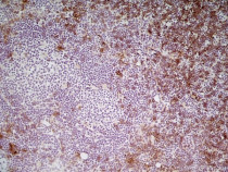
Immunohistochemistry: Mouse lymph node cryosection stained with ARG22899 anti-CD8 antibody [YTS105.18] followed by HRP conjugated Goat anti Rat IgG for detection. (Medium power).
-
ARG22899 anti-CD8 antibody [YTS105.18] IHC-Fr image
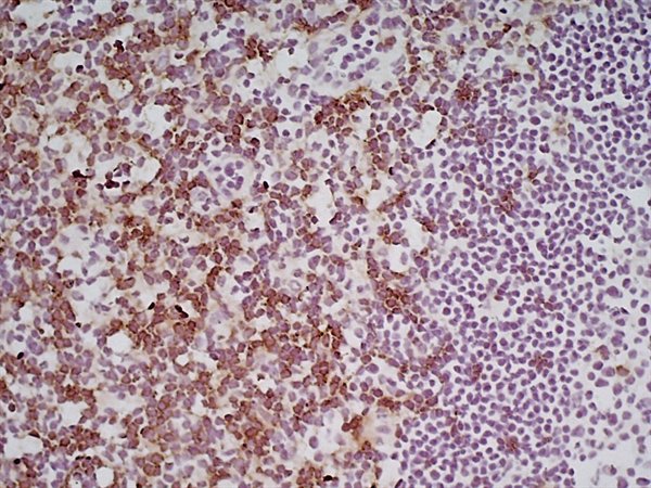
Immunohistochemistry: Mouse lymph node cryosection stained with ARG22899 anti-CD8 antibody [YTS105.18] followed by HRP conjugated Goat anti Rat IgG for detection. (High power).
-

ARG22899 anti-CD8 antibody [YTS105.18] IHC-Fr image
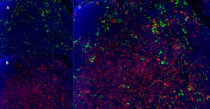
Immunohistochemistry: Mouse lymph node cryosection stained with Rat anti Mouse CD11b, clone 5C6, green in A and ARG22899 anti-CD8 antibody [YTS105.18], red in B. C is the merged image with nuclei counterstained blue using DAPI. (Low power).
-
ARG22899 anti-CD8 antibody [YTS105.18] IHC-Fr image
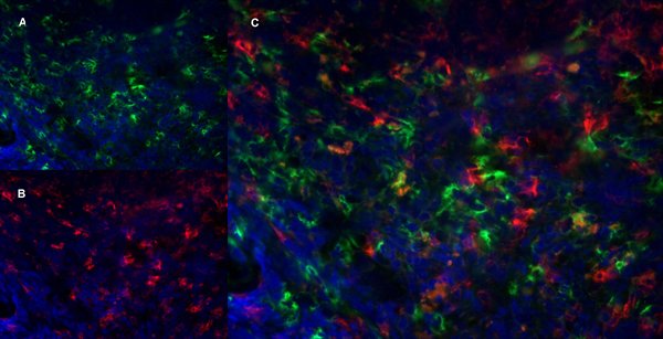
Immunohistochemistry: Mouse lymph node cryosection stained with Rat anti Mouse CD11b, clone 5C6, green in A and ARG22899 anti-CD8 antibody [YTS105.18], red in B. C is the merged image with nuclei counterstained blue using DAPI. (High power).
-

ARG22899 anti-CD8 antibody [YTS105.18] IHC-Fr image
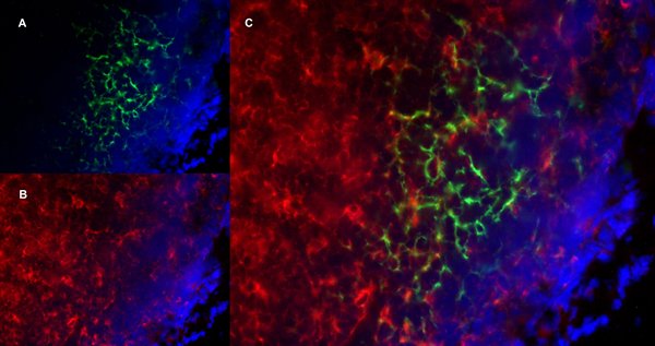
Immunohistochemistry: Mouse lymph node cryosection stained with Rat anti Mouse Ly-6B.2 antibody, clone 7/4, green in A and ARG22899 anti-CD8 antibody [YTS105.18], red in B. C is the merged image with nuclei counterstained blue using DAPI. (High power).
-

ARG22899 anti-CD8 antibody [YTS105.18] IHC-Fr image
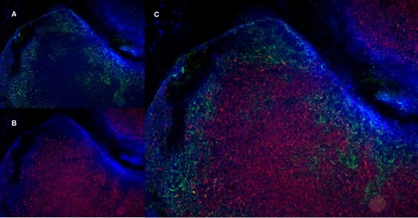
Immunohistochemistry: Mouse lymph node cryosection stained with Rat anti mouse CD19, clone 6D5, green in A and ARG22899 anti-CD8 antibody [YTS105.18], red in B. Merged image in C wih nuclei counterstained blue using DAPI. (Low power).
-

ARG22899 anti-CD8 antibody [YTS105.18] IHC-Fr image
Immunohistochemistry: Mouse lymph node cryosection stained with Rat anti mouse CD19, clone 6D5, green in A and ARG22899 anti-CD8 antibody [YTS105.18], red in B. Merged image in C wih nuclei counterstained blue using DAPI. (High power).