ARG22867
anti-CD8 antibody [CC63] (FITC)
anti-CD8 antibody [CC63] (FITC) for Flow cytometry and Bovine,Goat,Sheep
Developmental Biology antibody; Immune System antibody; Cytotoxic T antibody; Cytotoxic T Cell Surface Study antibody; Tumor-infiltrating Lymphocyte Study antibody
Overview
| Product Description | FITC-conjugated Mouse Monoclonal antibody [CC63] recognizes CD8 Mouse anti Bovine CD8 antibody, clone CC63 reacts with the bovine CD8 antigen expressed by a subset of T lymphocytes. The antibody precipitates molecules of 34kD and 38kD under reducing conditions. Clone CC63 has been reported as being suitable for use on formalin dichromate (FD5) fixed paraffin embedded tissue with amplification and antigen retrieval techniques (Gutierrez et al. 1999). |
|---|---|
| Tested Reactivity | Bov, Goat, Sheep |
| Tested Application | FACS |
| Host | Mouse |
| Clonality | Monoclonal |
| Clone | CC63 |
| Isotype | IgG2a |
| Target Name | CD8 |
| Antigen Species | Bovine |
| Conjugation | FITC |
| Alternate Names | T-cell surface glycoprotein CD8 alpha chain; Leu2; p32; T-lymphocyte differentiation antigen T8/Leu-2; CD8; MAL; CD antigen CD8a |
Application Instructions
| Application Suggestion |
|
||||
|---|---|---|---|---|---|
| Application Note | FACS: Use 10 µl of the suggested working dilution to label 10^6 cells in 100 µl. * The dilutions indicate recommended starting dilutions and the optimal dilutions or concentrations should be determined by the scientist. |
Properties
| Form | Liquid |
|---|---|
| Purification | Purification with Protein G. |
| Buffer | PBS, 0.09% Sodium azide and 1% BSA |
| Preservative | 0.09% Sodium azide |
| Stabilizer | 1% BSA |
| Concentration | 0.1 mg/ml |
| Storage Instruction | Aliquot and store in the dark at 2-8°C. Keep protected from prolonged exposure to light. Avoid repeated freeze/thaw cycles. Suggest spin the vial prior to opening. The antibody solution should be gently mixed before use. |
| Note | For laboratory research only, not for drug, diagnostic or other use. |
Bioinformation
| Database Links |
Swiss-port # P31783 Bovine T-cell surface glycoprotein CD8 alpha chain |
|---|---|
| Gene Symbol | CD8A |
| Gene Full Name | CD8a molecule |
| Background | CD8 antigen is a cell surface glycoprotein found on most cytotoxic T lymphocytes that mediates efficient cell-cell interactions within the immune system. The CD8 antigen acts as a coreceptor with the T-cell receptor on the T lymphocyte to recognize antigens displayed by an antigen presenting cell in the context of class I MHC molecules. The coreceptor functions as either a homodimer composed of two alpha chains or as a heterodimer composed of one alpha and one beta chain. Both alpha and beta chains share significant homology to immunoglobulin variable light chains. This gene encodes the CD8 alpha chain. Multiple transcript variants encoding different isoforms have been found for this gene. [provided by RefSeq, Nov 2011] |
| Function | CD8 is an integral membrane glycoprotein that plays an essential role in the immune response and serves multiple functions in responses against both external and internal offenses. In T-cells, functions primarily as a coreceptor for MHC class I molecule:peptide complex. The antigens presented by class I peptides are derived from cytosolic proteins while class II derived from extracellular proteins. Interacts simultaneously with the T-cell receptor (TCR) and the MHC class I proteins presented by antigen presenting cells (APCs). In turn, recruits the Src kinase LCK to the vicinity of the TCR-CD3 complex. LCK then initiates different intracellular signaling pathways by phosphorylating various substrates ultimately leading to lymphokine production, motility, adhesion and activation of cytotoxic T-lymphocytes (CTLs). This mechanism enables CTLs to recognize and eliminate infected cells and tumor cells. In NK-cells, the presence of CD8A homodimers at the cell surface provides a survival mechanism allowing conjugation and lysis of multiple target cells. CD8A homodimer molecules also promote the survival and differentiation of activated lymphocytes into memory CD8 T-cells. [UniProt] |
| Highlight | Related products: CD8 antibodies; CD8 ELISA Kits; CD8 Duos / Panels; Anti-Mouse IgG secondary antibodies; Related news: New antibody panels and duos for Tumor immune microenvironment Tumor-Infiltrating Lymphocytes (TILs) Detecting exosomal HMGB1 for ICD research |
| Research Area | Developmental Biology antibody; Immune System antibody; Cytotoxic T antibody; Cytotoxic T Cell Surface Study antibody; Tumor-infiltrating Lymphocyte Study antibody |
| Calculated MW | 26 kDa |
| PTM | All of the five most C-terminal cysteines form inter-chain disulfide bonds in dimers and higher multimers, while the four N-terminal cysteines do not. |
Images (3) Click the Picture to Zoom In
-
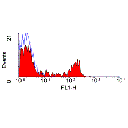
ARG22867 anti-CD8 antibody [CC63] (FITC) FACS image
Flow Cytometry: Bovine peripheral blood lymphocytes stained with ARG22867 anti-CD8 antibody [CC63] (FITC).
-
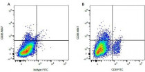
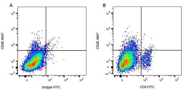
ARG22867 anti-CD8 antibody [CC63] (FITC) FACS image
Flow Cytometry: Figure A. A647 conjugated mouse anti bovine CD26 and FITC conjugated mouse IgG2a isotype control. Figure B. A647 conjugated mouse anti bovine CD26 and ARG22867 anti-CD8 antibody [CC63] (FITC). All experiments performed on red cell lysed bovine blood gated on mononuclear cells.
-

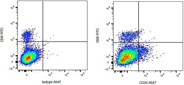
ARG22867 anti-CD8 antibody [CC63] (FITC) FACS image
Flow Cytometry: Figure A. ARG22867 anti-CD8 antibody [CC63] (FITC) and A647 conjugated mouse IgG1 isotype control. Figure B. ARG22867 anti-CD8 antibody [CC63] (FITC) and A647 conjugated mouse anti bovine CD26. All experiments performed on red cell lysed bovine blood gated on mononuclear cells.