ARG41758
anti-CD8 / CD8 alpha antibody
anti-CD8 / CD8 alpha antibody for Flow cytometry,IHC-Formalin-fixed paraffin-embedded sections,Western blot and Mouse,Rat
Developmental Biology antibody; Immune System antibody; Cytotoxic T antibody; Cytotoxic T Cell Surface Study antibody; Tumor-infiltrating Lymphocyte Study antibody
Overview
| Product Description | Rabbit Polyclonal antibody recognizes CD8 / CD8 alpha |
|---|---|
| Tested Reactivity | Ms, Rat |
| Tested Application | FACS, IHC-P, WB |
| Host | Rabbit |
| Clonality | Polyclonal |
| Isotype | IgG |
| Target Name | CD8 / CD8 alpha |
| Antigen Species | Human |
| Immunogen | Recombinant protein corresponding to K28-Y196 of Mouse CD8 / CD8 alpha. |
| Conjugation | Un-conjugated |
| Alternate Names | T-cell surface glycoprotein CD8 alpha chain; Leu2; p32; T-lymphocyte differentiation antigen T8/Leu-2; CD8; MAL; CD antigen CD8a |
Application Instructions
| Application Suggestion |
|
||||||||
|---|---|---|---|---|---|---|---|---|---|
| Application Note | IHC-P: Antigen Retrieval: Steam tissue section in Citrate buffer (pH 6.0) for 20 min followed by cooling at RT. * The dilutions indicate recommended starting dilutions and the optimal dilutions or concentrations should be determined by the scientist. |
||||||||
| Observed Size | ~ 36 kDa (26-40 kDa depending on glycosylation level) |
Properties
| Form | Liquid |
|---|---|
| Purification | Affinity purification with immunogen. |
| Buffer | 0.2% Na2HPO4, 0.9% NaCl, 0.05% Sodium azide and 4% Trehalose. |
| Preservative | 0.05% Sodium azide |
| Stabilizer | 4% Trehalose |
| Concentration | 0.5 mg/ml |
| Storage Instruction | For continuous use, store undiluted antibody at 2-8°C for up to a week. For long-term storage, aliquot and store at -20°C or below. Storage in frost free freezers is not recommended. Avoid repeated freeze/thaw cycles. Suggest spin the vial prior to opening. The antibody solution should be gently mixed before use. |
| Note | For laboratory research only, not for drug, diagnostic or other use. |
Bioinformation
| Database Links |
Swiss-port # P01731 Mouse T-cell surface glycoprotein CD8 alpha chain Swiss-port # P07725 Rat T-cell surface glycoprotein CD8 alpha chain |
|---|---|
| Gene Symbol | CD8A |
| Gene Full Name | CD8a molecule |
| Background | CD8 antigen is a cell surface glycoprotein found on most cytotoxic T lymphocytes that mediates efficient cell-cell interactions within the immune system. The CD8 antigen acts as a coreceptor with the T-cell receptor on the T lymphocyte to recognize antigens displayed by an antigen presenting cell in the context of class I MHC molecules. The coreceptor functions as either a homodimer composed of two alpha chains or as a heterodimer composed of one alpha and one beta chain. Both alpha and beta chains share significant homology to immunoglobulin variable light chains. This gene encodes the CD8 alpha chain. Multiple transcript variants encoding different isoforms have been found for this gene. [provided by RefSeq, Nov 2011] |
| Function | CD8 is an integral membrane glycoprotein that plays an essential role in the immune response and serves multiple functions in responses against both external and internal offenses. In T-cells, functions primarily as a coreceptor for MHC class I molecule:peptide complex. The antigens presented by class I peptides are derived from cytosolic proteins while class II derived from extracellular proteins. Interacts simultaneously with the T-cell receptor (TCR) and the MHC class I proteins presented by antigen presenting cells (APCs). In turn, recruits the Src kinase LCK to the vicinity of the TCR-CD3 complex. LCK then initiates different intracellular signaling pathways by phosphorylating various substrates ultimately leading to lymphokine production, motility, adhesion and activation of cytotoxic T-lymphocytes (CTLs). This mechanism enables CTLs to recognize and eliminate infected cells and tumor cells. In NK-cells, the presence of CD8A homodimers at the cell surface provides a survival mechanism allowing conjugation and lysis of multiple target cells. CD8A homodimer molecules also promote the survival and differentiation of activated lymphocytes into memory CD8 T-cells. [UniProt] |
| Cellular Localization | Isoform 1: Cell membrane; Single-pass type I membrane protein. Note=CD8A localizes to lipid rafts only when associated with its partner CD8B. Isoform 2: Secreted. [UniProt] |
| Highlight | Related products: CD8 antibodies; CD8 ELISA Kits; CD8 Duos / Panels; Anti-Rabbit IgG secondary antibodies; Related news: New antibody panels and duos for Tumor immune microenvironment Tumor-Infiltrating Lymphocytes (TILs) Detecting exosomal HMGB1 for ICD research |
| Research Area | Developmental Biology antibody; Immune System antibody; Cytotoxic T antibody; Cytotoxic T Cell Surface Study antibody; Tumor-infiltrating Lymphocyte Study antibody |
| Calculated MW | 26 kDa |
| PTM | All of the five most C-terminal cysteines form inter-chain disulfide bonds in dimers and higher multimers, while the four N-terminal cysteines do not. [UniProt] |
Images (4) Click the Picture to Zoom In
-
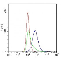
ARG41758 anti-CD8 / CD8 alpha antibody FACS image
Flow Cytometry: Mouse EL-4 cells stained with ARG41758 anti-CD8 / CD8 alpha antibody (blue) at 1 µg/10^6 cells. Isotype control (green). Cells alone (red).
-
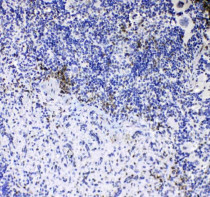
ARG41758 anti-CD8 / CD8 alpha antibody IHC-P image
Immunohistochemistry: Formalin-fixed and paraffin-embedded Mouse spleen tissue. Antigen Retrieval: Steam tissue section in Citrate buffer (pH 6.0) for 20 min followed by cooling at RT. The tissue section was stained with ARG41758 anti-CD8 / CD8 alpha antibody at 1 µg/ml dilution.
-

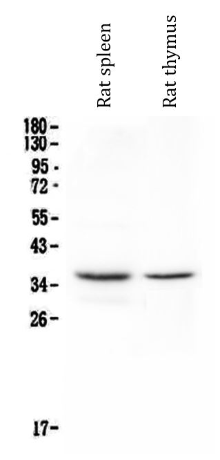
ARG41758 anti-CD8 / CD8 alpha antibody WB image
Western blot: Rat spleen and Rat thymus lysates stained with ARG41758 anti-CD8 / CD8 alpha antibody at 0.5 µg/ml dilution.
-
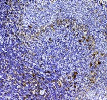
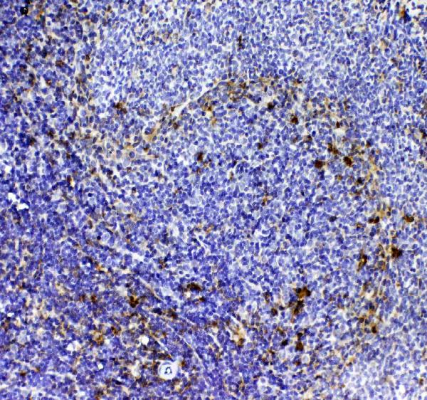
ARG41758 anti-CD8 / CD8 alpha antibody IHC-P image
Immunohistochemistry: Formalin-fixed and paraffin-embedded Rat spleen tissue. Antigen Retrieval: Steam tissue section in Citrate buffer (pH 6.0) for 20 min followed by cooling at RT. The tissue section was stained with ARG41758 anti-CD8 / CD8 alpha antibody at 1 µg/ml dilution.