ARG22839
anti-CD68 antibody [FA-11]
anti-CD68 antibody [FA-11] for CyTOF®-candidate,Flow cytometry,ICC/IF,IHC-Frozen sections,IHC-Formalin-fixed paraffin-embedded sections,Immunoprecipitation,Western blot and Mouse
Immune System antibody; Activated Macrophage/Microglia Study antibody; Neuroinflammation Study antibody; Active macroglial Marker antibody; M1/M2/TAM Marker antibody; Macrophage Marker antibody; M1 macrophage Marker antibody; Inflammatory Cell Marker antibody
Overview
| Product Description | Rat Monoclonal antibody [FA-11] recognizes CD68 CD68 antibody, clone FA-11Â recognizes mouse macrosialin which is a homolog of human CD68, which is classified as a unique scavenger receptor (ScR) family member. CD68 is considered a pan macrophage marker expressed on the intracellular lysosomes of tissue macrophages. |
|---|---|
| Tested Reactivity | Ms |
| Tested Application | CyTOF®-candidate, FACS, ICC/IF, IHC-Fr, IHC-P, IP, WB |
| Host | Rat |
| Clonality | Monoclonal |
| Clone | FA-11 |
| Isotype | IgG2a |
| Target Name | CD68 |
| Antigen Species | Mouse |
| Immunogen | Purified Concanavalin A acceptor glycoprotein from P815 cell line. |
| Conjugation | Un-conjugated |
| Alternate Names | Macrosialin; CD antigen CD68; LAMP4; Gp110; GP110; SCARD1 |
Application Instructions
| Application Suggestion |
|
||||||||||||||||
|---|---|---|---|---|---|---|---|---|---|---|---|---|---|---|---|---|---|
| Application Note | FACS: Membrane permeabilisation is required for this application. Use 10 µl of the suggested working dilution to label 10^6 cells in 100 µl. IHC-P: Antigen Retrieval: Boil tissue section in Sodium citrate buffer (pH 6.0). Staining has also been achieved without antigen retrieval. WB: Non-reducing conditions recommended. * The dilutions indicate recommended starting dilutions and the optimal dilutions or concentrations should be determined by the scientist. |
Properties
| Form | Liquid |
|---|---|
| Purification | Purification with Protein G. |
| Buffer | PBS and 0.09% Sodium azide |
| Preservative | 0.09% Sodium azide |
| Concentration | 1 mg/ml |
| Storage Instruction | For continuous use, store undiluted antibody at 2-8°C for up to a week. For long-term storage, aliquot and store at -20°C or below. Storage in frost free freezers is not recommended. Avoid repeated freeze/thaw cycles. Suggest spin the vial prior to opening. The antibody solution should be gently mixed before use. |
| Note | For laboratory research only, not for drug, diagnostic or other use. |
Bioinformation
| Database Links | |
|---|---|
| Gene Symbol | Cd68 |
| Gene Full Name | CD68 antigen |
| Background | CD68 is a 110-kD transmembrane glycoprotein that is highly expressed by human monocytes and tissue macrophages. It is a member of the lysosomal/endosomal-associated membrane glycoprotein (LAMP) family. The protein primarily localizes to lysosomes and endosomes with a smaller fraction circulating to the cell surface. It is a type I integral membrane protein with a heavily glycosylated extracellular domain and binds to tissue- and organ-specific lectins or selectins. The protein is also a member of the scavenger receptor family. Scavenger receptors typically function to clear cellular debris, promote phagocytosis, and mediate the recruitment and activation of macrophages. Alternative splicing results in multiple transcripts encoding different isoforms. [provided by RefSeq, Jul 2008] |
| Function | CD68 could play a role in phagocytic activities of tissue macrophages, both in intracellular lysosomal metabolism and extracellular cell-cell and cell-pathogen interactions. Binds to tissue- and organ-specific lectins or selectins, allowing homing of macrophage subsets to particular sites. Rapid recirculation of CD68 from endosomes and lysosomes to the plasma membrane may allow macrophages to crawl over selectin-bearing substrates or other cells. [UniProt] |
| Highlight | Related products: CD68 antibodies; CD68 Duos / Panels; Anti-Rat IgG secondary antibodies; Related news: CyTOF-candidate Antibodies New antibody panels and duos for Tumor immune microenvironment Tumor-Infiltrating Lymphocytes (TILs) Exploring Antiviral Immune Response Anti-SerpinB9 therapy, a new strategy for cancer therapy RIP1 activation and pathogenesis of NASH |
| Research Area | Immune System antibody; Activated Macrophage/Microglia Study antibody; Neuroinflammation Study antibody; Active macroglial Marker antibody; M1/M2/TAM Marker antibody; Macrophage Marker antibody; M1 macrophage Marker antibody; Inflammatory Cell Marker antibody |
| Calculated MW | 37 kDa |
| PTM | N- and O-glycosylated. |
Images (9) Click the Picture to Zoom In
-
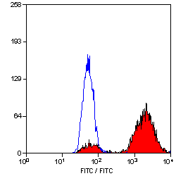
ARG22839 anti-CD68 antibody [FA-11] FACS image
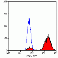
Flow Cytometry: Mouse peritoneal macrophages cells stained with ARG22839 anti-CD68 antibody [FA-11] following permeablisation with Leucoperm, visualised with Mouse adsorbed FITC conjugated F(ab')2 Goat anti Rat IgG.
-
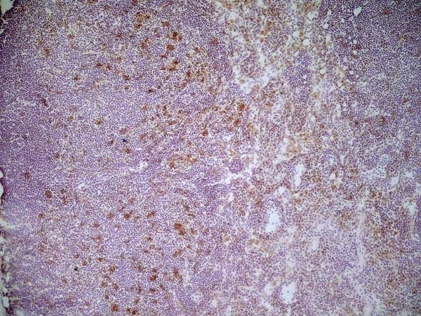
ARG22839 anti-CD68 antibody [FA-11] IHC-Fr image
Immunohistochemistry: Mouse spleen cryosection stained with ARG22839 anti-CD68 antibody [FA-11] followed by HRP-conjugated Goat anti Rat IgG.
-
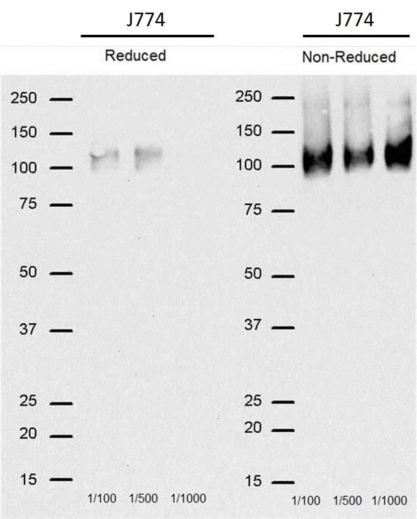
ARG22839 anti-CD68 antibody [FA-11] WB image
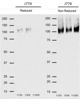
Western blot: J774 cells stained with ARG22839 anti-CD68 antibody [FA-11], under reducing and non-reducing condition.
-
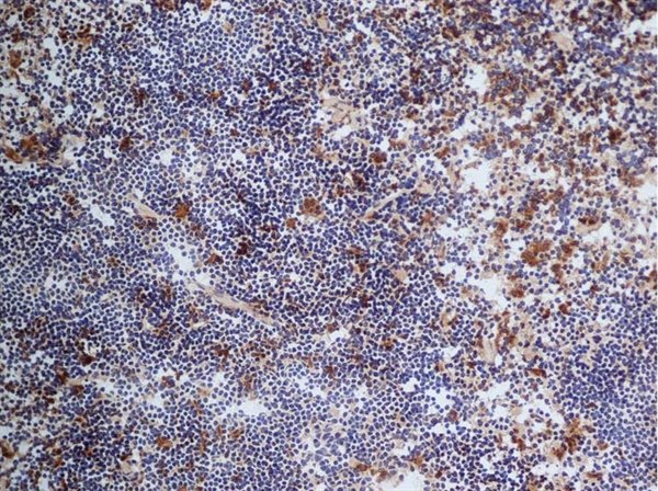
ARG22839 anti-CD68 antibody [FA-11] IHC-Fr image
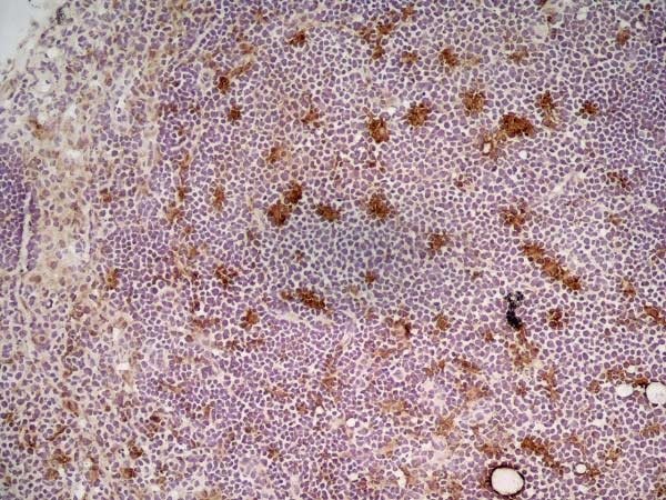
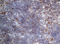
Immunohistochemistry: Frozen section of Mouse lymph node stained with ARG22839 anti-CD68 antibody [FA-11] at 1:100 dilution followed by HRP-conjugated Goat anti-Rat IgG at 1:50 dilution.
-

ARG22839 anti-CD68 antibody [FA-11] IHC-Fr image
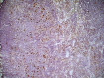
Immunohistochemistry: Frozen section of Mouse lymph node stained with ARG22839 anti-CD68 antibody [FA-11] at 1:100 dilution followed by HRP-conjugated Goat anti-Rat IgG at 1:50 dilution.
-

ARG22839 anti-CD68 antibody [FA-11] IHC-Fr image
Immunohistochemistry: Mouse lymph node cryosection stained with ARG22839 anti-CD68 antibody [FA-11] followed by Goat anti Rat IgG antibody. (High power).
-
ARG22839 anti-CD68 antibody [FA-11] IHC-Fr image
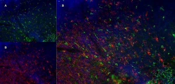
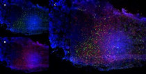
Immunohistochemistry: Mouse lymph node cryosection stained with ARG22839 anti-CD68 antibody [FA-11], green in A and Rat anti Mouse CD8 antibody, clone YTS105.18, red in B. C is the merged image with nuclei counterstained blue using DAPI. (Low power).
-
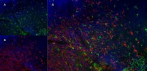
ARG22839 anti-CD68 antibody [FA-11] IHC-Fr image
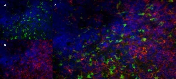
Immunohistochemistry: Mouse lymph node cryosection stained with ARG22839 anti-CD68 antibody [FA-11], green in A and Rat anti Mouse CD8 antibody, clone YTS105.18, red in B. C is the merged image with nuclei counterstained blue using DAPI. (Medium power).
-

ARG22839 anti-CD68 antibody [FA-11] IHC-Fr image
Immunohistochemistry: Mouse lymph node cryosection stained with ARG22839 anti-CD68 antibody [FA-11], green in A and Rat anti Mouse CD8 antibody, clone YTS105.18, red in B. C is the merged image with nuclei counterstained blue using DAPI. (High power).