ARG23387
anti-CD68 antibody [ED1]
anti-CD68 antibody [ED1] for Flow cytometry,IHC-Frozen sections,IHC-Formalin-fixed paraffin-embedded sections,Immunoprecipitation,Western blot and Mouse,Rat,Bovine
Immune System antibody; Activated Macrophage/Microglia Study antibody; Neuroinflammation Study antibody; Active macroglial Marker antibody; M1/M2/TAM Marker antibody; Macrophage Marker antibody; M1 macrophage Marker antibody; Inflammatory Cell Marker antibody

Overview
| Product Description | Mouse Monoclonal antibody [ED1] recognizes CD68 Mouse anti rat CD68, clone ED1 recognizes the rat ED1 antigen. The ED1 antigen is expressed on most macrophages populations, as well as on monocytes and is considered as a pan-macrophage marker in the rat. |
|---|---|
| Tested Reactivity | Ms, Rat, Bov |
| Species Does Not React With | Hrs |
| Tested Application | FACS, IHC-Fr, IHC-P, IP, WB |
| Host | Mouse |
| Clonality | Monoclonal |
| Clone | ED1 |
| Isotype | IgG1 |
| Target Name | CD68 |
| Antigen Species | Rat |
| Immunogen | Rat spleen cells |
| Conjugation | Un-conjugated |
| Alternate Names | Macrosialin; CD antigen CD68; LAMP4; Gp110; GP110; SCARD1 |
Application Instructions
| Application Suggestion |
|
||||||||||||
|---|---|---|---|---|---|---|---|---|---|---|---|---|---|
| Application Note | FACS: Membrane permeabilisation is required for this application. Use 10 µl of the suggested working dilution to label 10^6 cells in 100 µl. IHC-P: This product requires protein digestion pre-treatment of paraffin sections e.g. trypsin or pronase. * The dilutions indicate recommended starting dilutions and the optimal dilutions or concentrations should be determined by the scientist. |
Properties
| Form | Liquid |
|---|---|
| Purification | Purification with Protein A. |
| Buffer | PBS and 0.09% Sodium azide. |
| Preservative | 0.09% Sodium azide |
| Concentration | 1 mg/ml |
| Storage Instruction | For continuous use, store undiluted antibody at 2-8°C for up to a week. For long-term storage, aliquot and store at -20°C or below. Storage in frost free freezers is not recommended. Avoid repeated freeze/thaw cycles. Suggest spin the vial prior to opening. The antibody solution should be gently mixed before use. |
| Note | For laboratory research only, not for drug, diagnostic or other use. |
Bioinformation
| Database Links | |
|---|---|
| Gene Symbol | CD68 |
| Gene Full Name | CD68 molecule |
| Background | CD68 is a 110-kD transmembrane glycoprotein that is highly expressed by human monocytes and tissue macrophages. It is a member of the lysosomal/endosomal-associated membrane glycoprotein (LAMP) family. The protein primarily localizes to lysosomes and endosomes with a smaller fraction circulating to the cell surface. It is a type I integral membrane protein with a heavily glycosylated extracellular domain and binds to tissue- and organ-specific lectins or selectins. The protein is also a member of the scavenger receptor family. Scavenger receptors typically function to clear cellular debris, promote phagocytosis, and mediate the recruitment and activation of macrophages. Alternative splicing results in multiple transcripts encoding different isoforms. [provided by RefSeq, Jul 2008] |
| Function | CD68 could play a role in phagocytic activities of tissue macrophages, both in intracellular lysosomal metabolism and extracellular cell-cell and cell-pathogen interactions. Binds to tissue- and organ-specific lectins or selectins, allowing homing of macrophage subsets to particular sites. Rapid recirculation of CD68 from endosomes and lysosomes to the plasma membrane may allow macrophages to crawl over selectin-bearing substrates or other cells. [UniProt] |
| Highlight | Related products: CD68 antibodies; CD68 Duos / Panels; Anti-Mouse IgG secondary antibodies; Related news: New antibody panels and duos for Tumor immune microenvironment Tumor-Infiltrating Lymphocytes (TILs) Exploring Antiviral Immune Response Anti-SerpinB9 therapy, a new strategy for cancer therapy RIP1 activation and pathogenesis of NASH |
| Research Area | Immune System antibody; Activated Macrophage/Microglia Study antibody; Neuroinflammation Study antibody; Active macroglial Marker antibody; M1/M2/TAM Marker antibody; Macrophage Marker antibody; M1 macrophage Marker antibody; Inflammatory Cell Marker antibody |
| Calculated MW | 37 kDa |
| PTM | N- and O-glycosylated. [UniProt] |
Images (8) Click the Picture to Zoom In
-

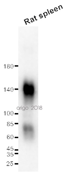
ARG23387 anti-CD68 antibody [ED1] WB image
Western blot: 20 µg of Rat spleen lysate stained with ARG23387 anti-CD68 antibody [ED1] at 1:500 dilution.
-
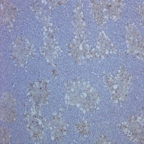
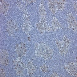
ARG23387 anti-CD68 antibody [ED1] IHC image
Immunohistochemistry: Rat liver, with induced hepatocellular damage, stained with ARG23387 anti-CD68 antibody [ED1].
-
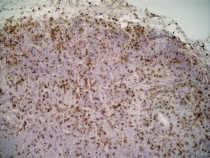
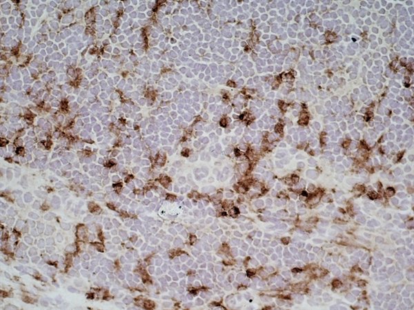
ARG23387 anti-CD68 antibody [ED1] IHC-Fr image
Immunohistochemistry: Rat lymph node cryosection stained with ARG23387 anti-CD68 antibody [ED1] followed by HRP-conjugated Goat anti Mouse IgG1 as a detection reagent. (Low power).
-
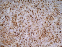
ARG23387 anti-CD68 antibody [ED1] IHC-Fr image
Immunohistochemistry: Rat lymph node cryosection stained with ARG23387 anti-CD68 antibody [ED1] followed by HRP-conjugated Goat anti Mouse IgG1 as a detection reagent. (Medium power).
-
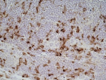
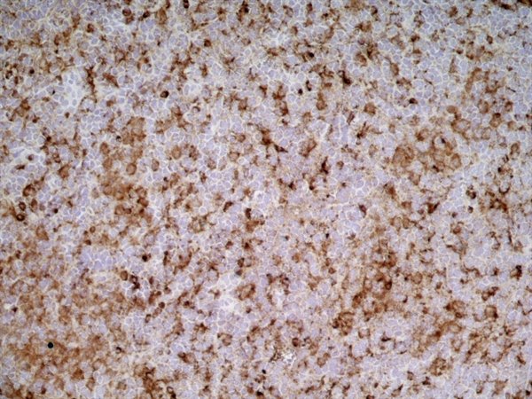
ARG23387 anti-CD68 antibody [ED1] IHC-Fr image
Immunohistochemistry: Rat lymph node cryosection stained with ARG23387 anti-CD68 antibody [ED1] followed by HRP-conjugated Goat anti Mouse IgG1 as a detection reagent. (High power).
-
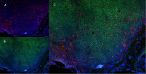
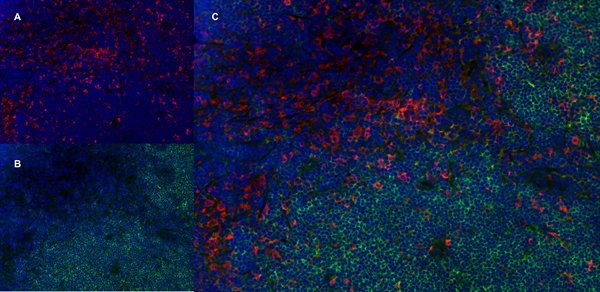
ARG23387 anti-CD68 antibody [ED1] IHC-Fr image
Immunohistochemistry: Rat lymph node cryosection stained with ARG23387 anti-CD68 antibody [ED1], red in A and Mouse anti Rat CD4, green in B. C is the merged image with nuclei counter-stained blue using DAPI. (Low power).
-
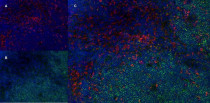
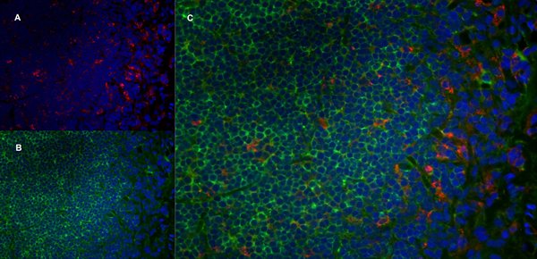
ARG23387 anti-CD68 antibody [ED1] IHC-Fr image
Immunohistochemistry: Rat lymph node cryosection stained with ARG23387 anti-CD68 antibody [ED1], red in A and Mouse anti Rat CD4, green in B. C is the merged image with nuclei counter-stained blue using DAPI. (Medium power).
-

ARG23387 anti-CD68 antibody [ED1] IHC-Fr image
Immunohistochemistry: Rat lymph node cryosection stained with ARG23387 anti-CD68 antibody [ED1], red in A and Mouse anti Rat CD4, green in B. C is the merged image with nuclei counter-stained blue using DAPI. (High power).


Customer's Feedback
Excellent
Review for anti-CD68 antibody [ED1]
Application:WB
Sample:Rat spleen
Sample Loading Amount:20 µg
Primary Antibody Dilution Factor:1:500
Primary Antibody Incubation Time:overnight
Primary Antibody Incubation Temperature:4 ºC

Specific References
Preparation and renoprotective effects of carboxymethyl chitosan oligosaccharide on adriamycin nephropathy
IHC-P / Rat


