ARG40200
anti-CD68 antibody
anti-CD68 antibody for IHC-Frozen sections,IHC-Formalin-fixed paraffin-embedded sections,Western blot and Mouse,Rat
Immune System antibody; Activated Macrophage/Microglia Study antibody; Neuroinflammation Study antibody; Active macroglial Marker antibody; M1/M2/TAM Marker antibody; Macrophage Marker antibody; M1 macrophage Marker antibody; Inflammatory Cell Marker antibody
Overview
| Product Description | Rabbit Polyclonal antibody recognizes CD68 |
|---|---|
| Tested Reactivity | Ms, Rat |
| Predict Reactivity | Hm |
| Tested Application | IHC-Fr, IHC-P, WB |
| Host | Rabbit |
| Clonality | Polyclonal |
| Isotype | IgG |
| Target Name | CD68 |
| Antigen Species | Mouse |
| Immunogen | Synthetic peptide corresponding to aa. 312-326 of Mouse CD68. (AFCITRRRQSTYQPL) |
| Conjugation | Un-conjugated |
| Alternate Names | Macrosialin; CD antigen CD68; LAMP4; Gp110; GP110; SCARD1 |
Application Instructions
| Application Suggestion |
|
||||||||
|---|---|---|---|---|---|---|---|---|---|
| Application Note | IHC-P: Antigen Retrieval: Heat mediation was performed in Citrate buffer (pH 6.0) for 20 min. * The dilutions indicate recommended starting dilutions and the optimal dilutions or concentrations should be determined by the scientist. |
Properties
| Form | Liquid |
|---|---|
| Purification | Affinity purification with immunogen. |
| Buffer | 0.9% NaCl, 0.2% Na2HPO4, 0.05% Sodium azide and 4% Trehalose. |
| Preservative | 0.05% Sodium azide |
| Stabilizer | 4% Trehalose |
| Concentration | 0.5 mg/ml |
| Storage Instruction | For continuous use, store undiluted antibody at 2-8°C for up to a week. For long-term storage, aliquot and store at -20°C or below. Storage in frost free freezers is not recommended. Avoid repeated freeze/thaw cycles. Suggest spin the vial prior to opening. The antibody solution should be gently mixed before use. |
| Note | For laboratory research only, not for drug, diagnostic or other use. |
Bioinformation
| Database Links | |
|---|---|
| Gene Symbol | CD68 |
| Gene Full Name | CD68 molecule |
| Background | CD68 is a 110-kD transmembrane glycoprotein that is highly expressed by human monocytes and tissue macrophages. It is a member of the lysosomal/endosomal-associated membrane glycoprotein (LAMP) family. The protein primarily localizes to lysosomes and endosomes with a smaller fraction circulating to the cell surface. It is a type I integral membrane protein with a heavily glycosylated extracellular domain and binds to tissue- and organ-specific lectins or selectins. The protein is also a member of the scavenger receptor family. Scavenger receptors typically function to clear cellular debris, promote phagocytosis, and mediate the recruitment and activation of macrophages. Alternative splicing results in multiple transcripts encoding different isoforms. [provided by RefSeq, Jul 2008] |
| Function | CD68 could play a role in phagocytic activities of tissue macrophages, both in intracellular lysosomal metabolism and extracellular cell-cell and cell-pathogen interactions. Binds to tissue- and organ-specific lectins or selectins, allowing homing of macrophage subsets to particular sites. Rapid recirculation of CD68 from endosomes and lysosomes to the plasma membrane may allow macrophages to crawl over selectin-bearing substrates or other cells. [UniProt] |
| Cellular Localization | Isoform Short: Cell membrane; Single-pass type I membrane protein. Isoform Long: Endosome membrane; Single-pass type I membrane protein. Lysosome membrane; Single-pass type I membrane protein. [UniProt] |
| Highlight | Related products: CD68 antibodies; CD68 Duos / Panels; Anti-Rabbit IgG secondary antibodies; Related news: New antibody panels and duos for Tumor immune microenvironment Tumor-Infiltrating Lymphocytes (TILs) Exploring Antiviral Immune Response Anti-SerpinB9 therapy, a new strategy for cancer therapy RIP1 activation and pathogenesis of NASH |
| Research Area | Immune System antibody; Activated Macrophage/Microglia Study antibody; Neuroinflammation Study antibody; Active macroglial Marker antibody; M1/M2/TAM Marker antibody; Macrophage Marker antibody; M1 macrophage Marker antibody; Inflammatory Cell Marker antibody |
| Calculated MW | 37 kDa |
| PTM | N- and O-glycosylated. [UniProt] |
Images (8) Click the Picture to Zoom In
-
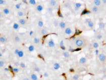
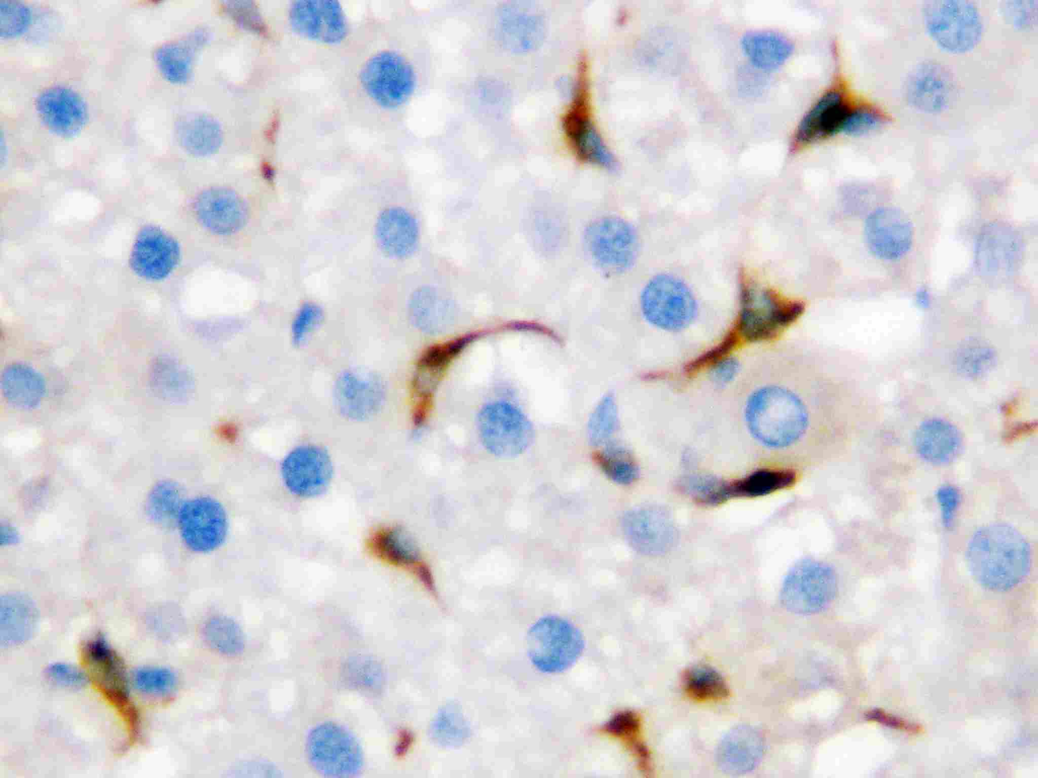
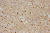
ARG40200 anti-CD68 antibody IHC-P image
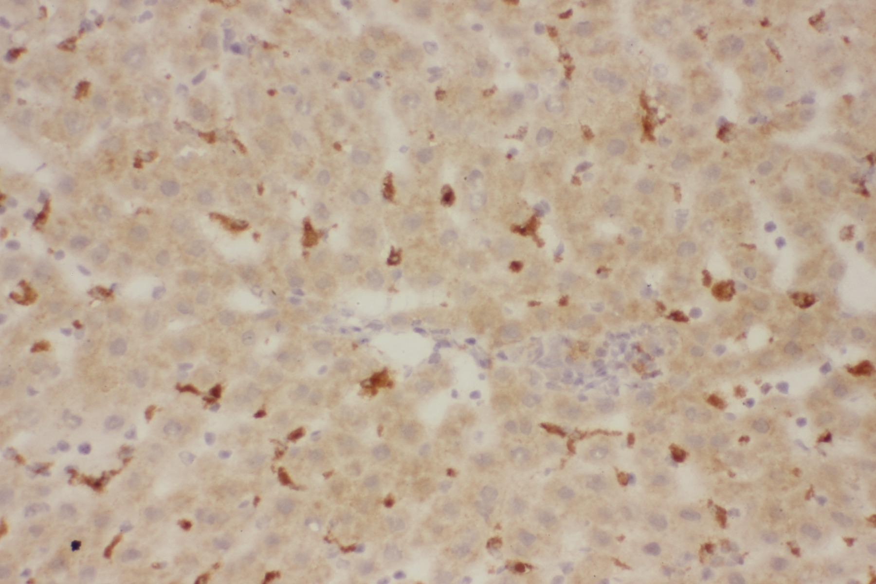
Immunohistochemistry: Paraffin-embedded Rat liver tissue. Antigen Retrieval: Heat mediation was performed in Citrate buffer (pH 6.0, epitope retrieval solution) for 20 min. The tissue section was blocked with 10% goat serum. The tissue section was then stained with ARG40200 anti-CD68 antibody at 1 µg/ml, overnight at 4°C.
-
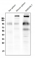
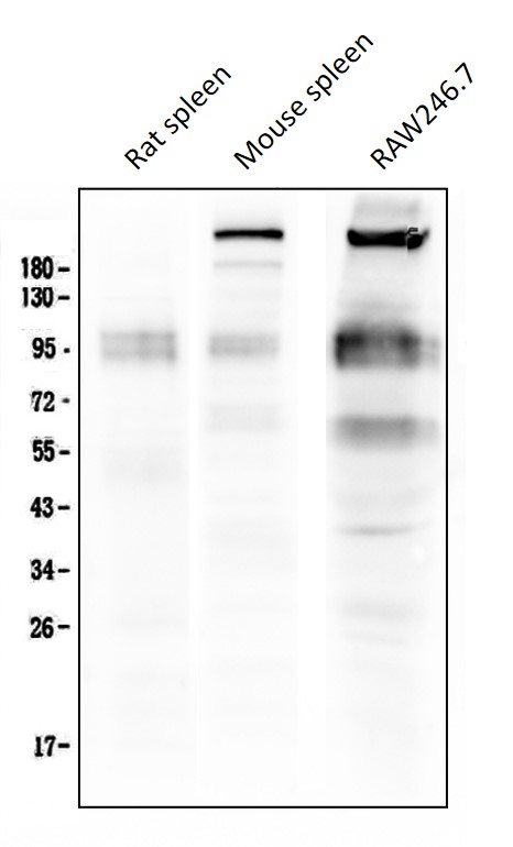
ARG40200 anti-CD68 antibody WB image
Western blot: 50 µg of samples under reducing conditions. Rat spleen, Mouse spleen and RAW246.7 cell lysates stained with ARG40200 anti-CD68 antibody at 0.5 µg/ml, overnight at 4°C.
-

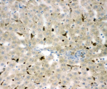
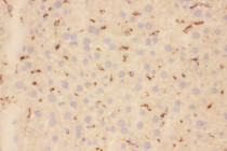
ARG40200 anti-CD68 antibody IHC-P image
Immunohistochemistry: Paraffin-embedded Rat liver tissue. Antigen Retrieval: Heat mediation was performed in Citrate buffer (pH 6.0, epitope retrieval solution) for 20 min. The tissue section was blocked with 10% goat serum. The tissue section was then stained with ARG40200 anti-CD68 antibody at 1 µg/ml, overnight at 4°C.
-
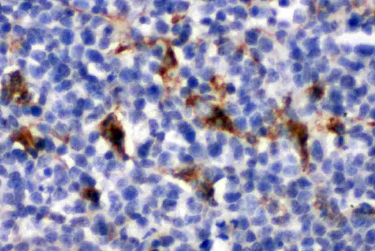
ARG40200 anti-CD68 antibody IHC-P image
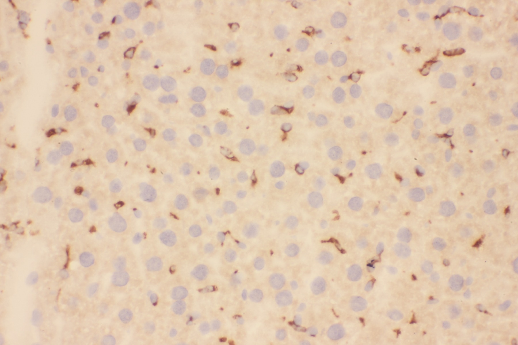
Immunohistochemistry: Paraffin-embedded Mouse spleen tissue. Antigen Retrieval: Heat mediation was performed in Citrate buffer (pH 6.0, epitope retrieval solution) for 20 min. The tissue section was blocked with 10% goat serum. The tissue section was then stained with ARG40200 anti-CD68 antibody at 1 µg/ml, overnight at 4°C.
-
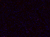
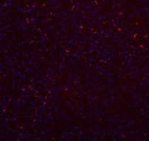
ARG40200 anti-CD68 antibody IHC-P image
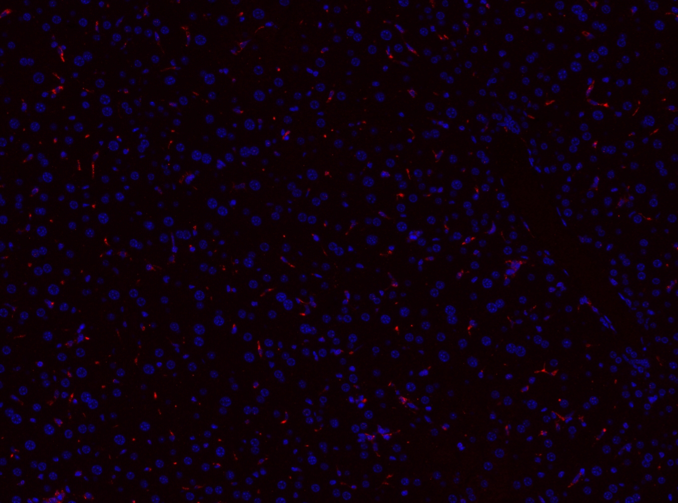
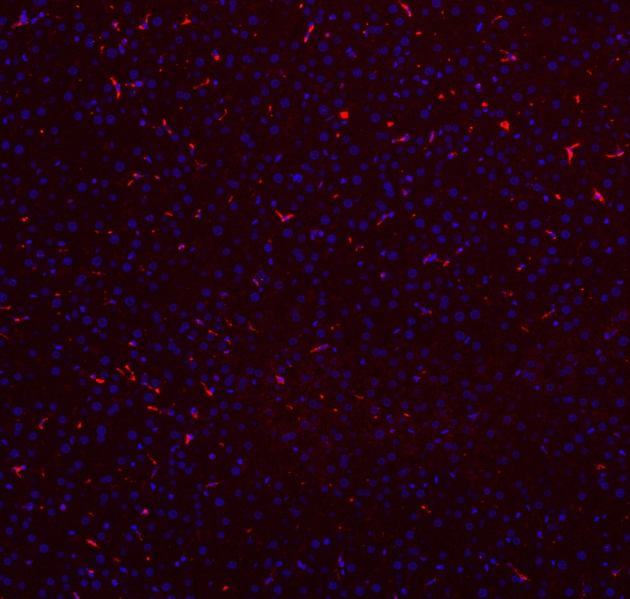
Immunohistochemistry: Paraffin-embedded Rat liver tissue. Antigen Retrieval: Heat mediation was performed in Citrate buffer (pH 6.0, epitope retrieval solution) for 20 min. The tissue section was blocked with 10% goat serum. The tissue section was then stained with ARG40200 anti-CD68 antibody (red) at 1 µg/ml, overnight at 4°C. The section was counterstained with DAPI (blue).
-
ARG40200 anti-CD68 antibody IHC-P image
Immunohistochemistry: Paraffin-embedded Rat liver tissue. Antigen Retrieval: Heat mediation was performed in Citrate buffer (pH 6.0, epitope retrieval solution) for 20 min. The tissue section was blocked with 10% goat serum. The tissue section was then stained with ARG40200 anti-CD68 antibody (red) at 1 µg/ml, overnight at 4°C. The section was counterstained with DAPI (blue).
-
ARG40200 anti-CD68 antibody IHC-Fr image
Immunohistochemistry: Frozen section of Rat liver tissue. The tissue section was blocked with 10% goat serum. The tissue section was then stained with ARG40200 anti-CD68 antibody at 1 μg/ml dilution, overnight at 4°C.
-
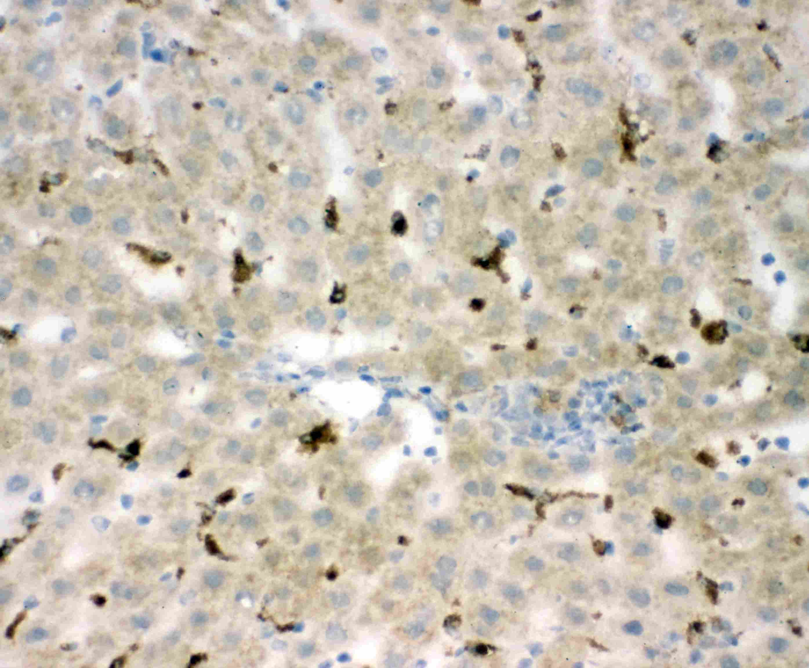
ARG40200 anti-CD68 antibody IHC-Fr image
Immunohistochemistry: Frozen section of Mouse liver tissue. The tissue section was blocked with 10% goat serum. The tissue section was then stained with ARG40200 anti-CD68 antibody at 1 μg/ml dilution, overnight at 4°C.