ARG53887
anti-CD63 antibody [MEM-259] (PE)
anti-CD63 antibody [MEM-259] (PE) for Flow cytometry and Human
Cancer antibody; Cell Biology and Cellular Response antibody; Controls and Markers antibody; Immune System antibody
Overview
| Product Description | PE-conjugated Mouse Monoclonal antibody [MEM-259] recognizes CD63 |
|---|---|
| Tested Reactivity | Hu |
| Tested Application | FACS |
| Specificity | The clone MEM-259 reacts with CD63 (LAMP-3), a 40-60 kDa tetraspan glycoprotein expressed by granulocytes, platelets, T cells, monocytes/macrophages and endothelial cells. Cell surface exposition of CD63 is usually activation-dependent. |
| Host | Mouse |
| Clonality | Monoclonal |
| Clone | MEM-259 |
| Isotype | IgG1 |
| Target Name | CD63 |
| Immunogen | HPB-ALL T cell line |
| Conjugation | PE |
| Alternate Names | Tspan-30; CD63 antigen; Tetraspanin-30; CD antigen CD63; Lysosomal-associated membrane protein 3; OMA81H; Ocular melanoma-associated antigen; Granulophysin; TSPAN30; Melanoma-associated antigen ME491; MLA1; LAMP-3; ME491 |
Application Instructions
| Application Suggestion |
|
||||
|---|---|---|---|---|---|
| Application Note | * The dilutions indicate recommended starting dilutions and the optimal dilutions or concentrations should be determined by the scientist. |
Properties
| Form | Liquid |
|---|---|
| Purification Note | The purified antibody is conjugated with R-Phycoerythrin (PE) under optimum conditions. The conjugate is purified by size-exclusion chromatography and adjusted for direct use. No reconstitution is necessary. |
| Buffer | PBS, 15 mM Sodium azide and 0.2% (w/v) high-grade protease free BSA |
| Preservative | 15 mM Sodium azide |
| Stabilizer | 0.2% (w/v) high-grade protease free BSA |
| Storage Instruction | Aliquot and store in the dark at 2-8°C. Keep protected from prolonged exposure to light. Avoid repeated freeze/thaw cycles. Suggest spin the vial prior to opening. The antibody solution should be gently mixed before use. |
| Note | For laboratory research only, not for drug, diagnostic or other use. |
Bioinformation
| Database Links | |
|---|---|
| Gene Symbol | CD63 |
| Gene Full Name | CD63 molecule |
| Background | CD63 (LAMP-3, lysosome-associated membrane protein-3), a glycoprotein of tetraspanin family, is present in late endosomes, lysosomes and secretory vesicles of various cell types. It is also present in the plasma membrane, usually following cell activation. Hence, it has become an widely used basophil activation marker. In mast cells, however, CD63 exposition does not need their activation. CD63 interacts with integrins and affects phagocytosis and cell migration, it is also involved in H/K-ATPase trafficking regulation of ROMK1 channels. CD63 also serves as a T-cell costimulation molecule. Expression of CD63 can be used for predicting the prognosis in earlier stages of carcinomas. |
| Function | Functions as cell surface receptor for TIMP1 and plays a role in the activation of cellular signaling cascades. Plays a role in the activation of ITGB1 and integrin signaling, leading to the activation of AKT, FAK/PTK2 and MAP kinases. Promotes cell survival, reorganization of the actin cytoskeleton, cell adhesion, spreading and migration, via its role in the activation of AKT and FAK/PTK2. Plays a role in VEGFA signaling via its role in regulating the internalization of KDR/VEGFR2. Plays a role in intracellular vesicular transport processes, and is required for normal trafficking of the PMEL luminal domain that is essential for the development and maturation of melanocytes. Plays a role in the adhesion of leukocytes onto endothelial cells via its role in the regulation of SELP trafficking. May play a role in mast cell degranulation in response to Ms4a2/FceRI stimulation, but not in mast cell degranulation in response to other stimuli. [UniProt] |
| Highlight | Related products: CD63 antibodies; Anti-Mouse IgG secondary antibodies; Related news: Tools for studying Exosomes |
| Research Area | Cancer antibody; Cell Biology and Cellular Response antibody; Controls and Markers antibody; Immune System antibody |
| Calculated MW | 26 kDa |
| PTM | Palmitoylated at a low, basal level in unstimulated platelets. The level of palmitoylation increases when platelets are activated by thrombin (in vitro). |
Images (1) Click the Picture to Zoom In
-
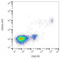
ARG53887 anti-CD63 antibody [MEM-259] (PE) FACS image
Flow Cytometry: IgE-activated peripheral blood stained with ARG53887 anti-CD63 antibody [MEM-259] (PE).

Clone References
The impact of disparate isolation methods for extracellular vesicles on downstream RNA profiling.
IHC / Human


Exosomes from hepatitis C infected patients transmit HCV infection and contain replication competent viral RNA in complex with Ago2-miR122-HSP90.
Investigation of soluble and transmembrane CTLA-4 isoforms in serum and microvesicles.
EM / Human
A gestational profile of placental exosomes in maternal plasma and their effects on endothelial cell migration.
WB / Human
Epstein-Barr virus-encoded small RNAs (EBERs) are present in fractions related to exosomes released by EBV-transformed cells.
WB / Human
A human blood-brain barrier transcytosis assay reveals antibody transcytosis influenced by pH-dependent receptor binding.
ICC/IF / Human
Syntenin-ALIX exosome biogenesis and budding into multivesicular bodies are controlled by ARF6 and PLD2.


