ARG22811
anti-CD4 antibody [YKIX302.9] (PE)
anti-CD4 antibody [YKIX302.9] (PE) for Flow cytometry and Dog
Developmental Biology antibody; Immune System antibody; Regulatory T cells Study antibody; T-cell infiltration Study antibody; Tumor-infiltrating Lymphocyte Study antibody
Overview
| Product Description | PE-conjugated Rat Monoclonal antibody [YKIX302.9] recognizes CD4 Rat anti Dog CD4 antibody, clone YKIX302.9, is a monoclonal antibody specific for the canine CD4 cell surface antigen. Clone YKIX302.9 was clustered at the first Canine Leukocyte Antigen Workshop (CLAW) [Cobbold et al. 1992] along with clone CA13.1E4. Rat anti Dog CD4 (YKIX302.9) has been demonstrated to partially deplete circulating T lymphocytes when administered in vivo, but alone was not sufficient to prolong allograft survival in a canine transplant model (Watson et al. 1993). Uniquely amongst mammals, canine CD4 is expressed by neutrophils as well as by lymphocytes subsets Moore et al. 1992. Rat anti Canine CD4 (YKIX302.9) forms part of a panel of anti canine monoclonal antibodies used extensively in the evaluation of leukemic status in dogs (Villiers 2002). |
|---|---|
| Tested Reactivity | Dog |
| Tested Application | FACS |
| Host | Rat |
| Clonality | Monoclonal |
| Clone | YKIX302.9 |
| Isotype | IgG2a |
| Target Name | CD4 |
| Antigen Species | Dog |
| Immunogen | Canine concanavilin A activated T cell blasts. |
| Conjugation | PE |
| Alternate Names | CD4mut; CD antigen CD4; T-cell surface glycoprotein CD4; T-cell surface antigen T4/Leu-3 |
Application Instructions
| Application Suggestion |
|
||||
|---|---|---|---|---|---|
| Application Note | FACS: Use 10 µl of the suggested working dilution to label 10^6 cells or 100 µl whole blood. * The dilutions indicate recommended starting dilutions and the optimal dilutions or concentrations should be determined by the scientist. |
Properties
| Form | Liquid |
|---|---|
| Purification | Purification with Protein G. |
| Buffer | PBS, 0.09% Sodium azide, 1% BSA and 5% Sucrose |
| Preservative | 0.09% Sodium azide |
| Stabilizer | 1% BSA and 5% Sucrose |
| Storage Instruction | Aliquot and store in the dark at 2-8°C. Keep protected from prolonged exposure to light. Avoid repeated freeze/thaw cycles. Suggest spin the vial prior to opening. The antibody solution should be gently mixed before use. |
| Note | For laboratory research only, not for drug, diagnostic or other use. |
Bioinformation
| Gene Symbol | CD4 |
|---|---|
| Gene Full Name | CD4 molecule |
| Background | CD4 is a membrane glycoprotein of T lymphocytes that interacts with major histocompatibility complex class II antigenes and is also a receptor for the human immunodeficiency virus. This gene is expressed not only in T lymphocytes, but also in B cells, macrophages, and granulocytes. It is also expressed in specific regions of the brain. The protein functions to initiate or augment the early phase of T-cell activation, and may function as an important mediator of indirect neuronal damage in infectious and immune-mediated diseases of the central nervous system. Multiple alternatively spliced transcript variants encoding different isoforms have been identified in this gene. [provided by RefSeq, Aug 2010] |
| Function | CD4 is an integral membrane glycoprotein that plays an essential role in the immune response and serves multiple functions in responses against both external and internal offenses. In T-cells, functions primarily as a coreceptor for MHC class II molecule:peptide complex. The antigens presented by class II peptides are derived from extracellular proteins while class I peptides are derived from cytosolic proteins. Interacts simultaneously with the T-cell receptor (TCR) and the MHC class II presented by antigen presenting cells (APCs). In turn, recruits the Src kinase LCK to the vicinity of the TCR-CD3 complex. LCK then initiates different intracellular signaling pathways by phosphorylating various substrates ultimately leading to lymphokine production, motility, adhesion and activation of T-helper cells. In other cells such as macrophages or NK cells, plays a role in differentiation/activation, cytokine expression and cell migration in a TCR/LCK-independent pathway. Participates in the development of T-helper cells in the thymus and triggers the differentiation of monocytes into functional mature macrophages. [UniProt] |
| Highlight | Related products: CD4 antibodies; CD4 ELISA Kits; CD4 Duos / Panels; Anti-Rat IgG secondary antibodies; Related news: New antibody panels and duos for Tumor immune microenvironment Tumor-Infiltrating Lymphocytes (TILs) |
| Research Area | Developmental Biology antibody; Immune System antibody; Regulatory T cells Study antibody; T-cell infiltration Study antibody; Tumor-infiltrating Lymphocyte Study antibody |
| Calculated MW | 51 kDa |
| PTM | Palmitoylation and association with LCK contribute to the enrichment of CD4 in lipid rafts. |
Images (2) Click the Picture to Zoom In
-
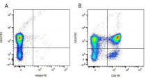
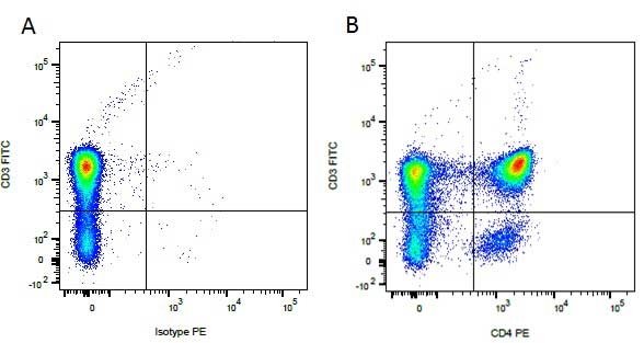
ARG22811 anti-CD4 antibody [YKIX302.9] (PE) FACS image
Flow Cytometry: Figure A. FITC conjugated Mouse anti Canine CD3 and PE-conjugated Rat IgG2a isotype control. Figure B. FITC conjugated Mouse anti Canine CD3 and ARG22811 anti-CD4 antibody [YKIX302.9] (PE). All experiments performed on red cell lysed canine blood gated on mononuclear cells.
-
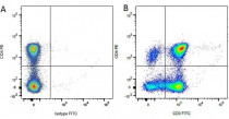
ARG22811 anti-CD4 antibody [YKIX302.9] (PE) FACS image
Flow Cytometry: Figure A. ARG22811 anti-CD4 antibody [YKIX302.9] (PE) and FITC conjugated Mouse IgG1 isotype control. Figure B. ARG22811 anti-CD4 antibody [YKIX302.9] (PE) and FITC conjugated Mouse anti Canine CD3. All experiments performed on red cell lysed canine blood gated on mononuclear cells.