ARG62837
anti-CD4 antibody [GK1.5] (FITC)
anti-CD4 antibody [GK1.5] (FITC) for Flow cytometry,ICC/IF,IHC-Frozen sections and Mouse
Developmental Biology antibody; Immune System antibody; Regulatory T cells Study antibody; T-cell infiltration Study antibody; Tumor-infiltrating Lymphocyte Study antibody
Overview
| Product Description | FITC-conjugated Rat Monoclonal antibody [GK1.5] recognizes CD4 |
|---|---|
| Tested Reactivity | Ms |
| Tested Application | FACS, ICC/IF, IHC-Fr |
| Specificity | The clone GK1.5 reacts with an extracellular epitope of mouse CD4 transmembrane glycoprotein (55 kDa). |
| Host | Rat |
| Clonality | Monoclonal |
| Clone | GK1.5 |
| Isotype | IgG2b, kappa |
| Target Name | CD4 |
| Antigen Species | Mouse |
| Immunogen | Mouse CTL clone V4 cells |
| Epitope | extracellular epitope of mouse CD4 |
| Conjugation | FITC |
| Alternate Names | CD4mut; CD antigen CD4; T-cell surface glycoprotein CD4; T-cell surface antigen T4/Leu-3 |
Application Instructions
| Application Suggestion |
|
||||||||
|---|---|---|---|---|---|---|---|---|---|
| Application Note | * The dilutions indicate recommended starting dilutions and the optimal dilutions or concentrations should be determined by the scientist. |
Properties
| Form | Liquid |
|---|---|
| Purification Note | The purified antibody is conjugated with Fluorescein isothiocyanate (FITC) under optimum conditions. The reagent is free of unconjugated FITC. |
| Buffer | PBS (pH 7.4) and 15 mM Sodium azide |
| Preservative | 15 mM Sodium azide |
| Concentration | 0.5 mg/ml |
| Storage Instruction | Aliquot and store in the dark at 2-8°C. Keep protected from prolonged exposure to light. Avoid repeated freeze/thaw cycles. Suggest spin the vial prior to opening. The antibody solution should be gently mixed before use. |
| Note | For laboratory research only, not for drug, diagnostic or other use. |
Bioinformation
| Database Links | |
|---|---|
| Gene Symbol | CD4 |
| Gene Full Name | CD4 molecule |
| Background | CD4 is a membrane glycoprotein of T lymphocytes that interacts with major histocompatibility complex class II antigenes and is also a receptor for the human immunodeficiency virus. This gene is expressed not only in T lymphocytes, but also in B cells, macrophages, and granulocytes. It is also expressed in specific regions of the brain. The protein functions to initiate or augment the early phase of T-cell activation, and may function as an important mediator of indirect neuronal damage in infectious and immune-mediated diseases of the central nervous system. Multiple alternatively spliced transcript variants encoding different isoforms have been identified in this gene. [provided by RefSeq, Aug 2010] |
| Function | CD4 is an integral membrane glycoprotein that plays an essential role in the immune response and serves multiple functions in responses against both external and internal offenses. In T-cells, functions primarily as a coreceptor for MHC class II molecule:peptide complex. The antigens presented by class II peptides are derived from extracellular proteins while class I peptides are derived from cytosolic proteins. Interacts simultaneously with the T-cell receptor (TCR) and the MHC class II presented by antigen presenting cells (APCs). In turn, recruits the Src kinase LCK to the vicinity of the TCR-CD3 complex. LCK then initiates different intracellular signaling pathways by phosphorylating various substrates ultimately leading to lymphokine production, motility, adhesion and activation of T-helper cells. In other cells such as macrophages or NK cells, plays a role in differentiation/activation, cytokine expression and cell migration in a TCR/LCK-independent pathway. Participates in the development of T-helper cells in the thymus and triggers the differentiation of monocytes into functional mature macrophages. [UniProt] |
| Highlight | Related products: CD4 antibodies; CD4 ELISA Kits; CD4 Duos / Panels; Anti-Rat IgG secondary antibodies; Related news: New antibody panels and duos for Tumor immune microenvironment Tumor-Infiltrating Lymphocytes (TILs) |
| Research Area | Developmental Biology antibody; Immune System antibody; Regulatory T cells Study antibody; T-cell infiltration Study antibody; Tumor-infiltrating Lymphocyte Study antibody |
| Calculated MW | 51 kDa |
| PTM | Palmitoylation and association with LCK contribute to the enrichment of CD4 in lipid rafts. |
Images (2) Click the Picture to Zoom In
-
-
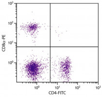
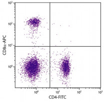
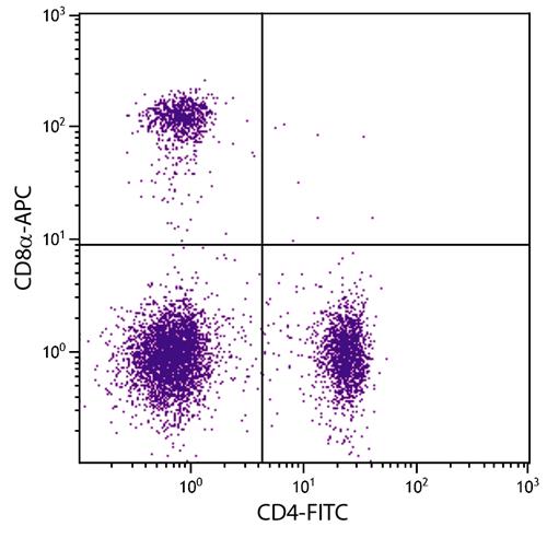
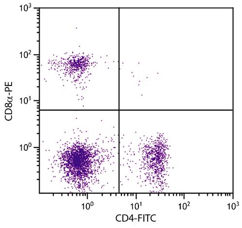
ARG62837 anti-CD4 antibody [GK1.5] (FITC) FACS image
Flow Cytometry: BALB/c Mouse splenocytes stained with ARG62837 anti-CD4 antibody [GK1.5] (FITC) and ARG62941 anti-CD8a antibody [53-6.7] (PE).
Clone References
Myxoma virus expressing a fusion protein of interleukin-15 (IL15) and IL15 receptor alpha has enhanced antitumor activity.
IHC-Fr / Mouse


Autologous tumor vaccine modified with recombinant new castle disease virus expressing IL-7 promotes antitumor immune response.
Supplementation of influenza split vaccines with conserved M2 ectodomains overcomes strain specificity and provides long-term cross protection.
A quantitative study of the mechanisms behind thymic atrophy in Gαi2-deficient mice during colitis development.
FACS / Mouse


