ARG23427
anti-CD4 antibody [CC8] (PE)
anti-CD4 antibody [CC8] (PE) for Flow cytometry and Bovine
Developmental Biology antibody; Immune System antibody; Regulatory T cells Study antibody; T-cell infiltration Study antibody; Tumor-infiltrating Lymphocyte Study antibody
Overview
| Product Description | PE-conjugated Mouse Monoclonal antibody [CC8] recognizes CD4 Mouse anti Bovine CD4 antibody, clone CC8 recognizes bovine CD4, the homolog of human CD4 and immunoprecipitates a ~50 kDa molecule. The phenotype, tissue distribution and function of T-cells expressing the bovine CD4 antigen are similar to those in other species. However, expression on macrophages has not yet been detected. Clone CC8 has been reported as being suitable for use on formalin dichromate (FD5) fixed paraffin embedded tissue with amplification and antigen retrieval techniques (Eskra et al. 1991). |
|---|---|
| Tested Reactivity | Bov |
| Tested Application | FACS |
| Host | Mouse |
| Clonality | Monoclonal |
| Clone | CC8 |
| Isotype | IgG2a |
| Target Name | CD4 |
| Antigen Species | Bovine |
| Immunogen | Bovine lymphocytes. |
| Conjugation | PE |
| Alternate Names | CD4mut; CD antigen CD4; T-cell surface glycoprotein CD4; T-cell surface antigen T4/Leu-3 |
Application Instructions
| Application Suggestion |
|
||||
|---|---|---|---|---|---|
| Application Note | FACS: Use 10 µl of the suggested working dilution to label 10^6 cells in 100 µl. * The dilutions indicate recommended starting dilutions and the optimal dilutions or concentrations should be determined by the scientist. |
Properties
| Form | Liquid |
|---|---|
| Purification | Purification with Protein A. |
| Buffer | PBS, 0.09% Sodium azide, 1% BSA and 5% Sucrose. |
| Preservative | 0.09% Sodium azide |
| Stabilizer | 1% BSA and 5% Sucrose |
| Storage Instruction | Aliquot and store in the dark at 2-8°C. Keep protected from prolonged exposure to light. Avoid repeated freeze/thaw cycles. Suggest spin the vial prior to opening. The antibody solution should be gently mixed before use. |
| Note | For laboratory research only, not for drug, diagnostic or other use. |
Bioinformation
| Gene Symbol | CD4 |
|---|---|
| Gene Full Name | CD4 molecule |
| Background | CD4 is a membrane glycoprotein of T lymphocytes that interacts with major histocompatibility complex class II antigenes and is also a receptor for the human immunodeficiency virus. This gene is expressed not only in T lymphocytes, but also in B cells, macrophages, and granulocytes. It is also expressed in specific regions of the brain. The protein functions to initiate or augment the early phase of T-cell activation, and may function as an important mediator of indirect neuronal damage in infectious and immune-mediated diseases of the central nervous system. Multiple alternatively spliced transcript variants encoding different isoforms have been identified in this gene. [provided by RefSeq, Aug 2010] |
| Function | CD4 is an integral membrane glycoprotein that plays an essential role in the immune response and serves multiple functions in responses against both external and internal offenses. In T-cells, functions primarily as a coreceptor for MHC class II molecule:peptide complex. The antigens presented by class II peptides are derived from extracellular proteins while class I peptides are derived from cytosolic proteins. Interacts simultaneously with the T-cell receptor (TCR) and the MHC class II presented by antigen presenting cells (APCs). In turn, recruits the Src kinase LCK to the vicinity of the TCR-CD3 complex. LCK then initiates different intracellular signaling pathways by phosphorylating various substrates ultimately leading to lymphokine production, motility, adhesion and activation of T-helper cells. In other cells such as macrophages or NK cells, plays a role in differentiation/activation, cytokine expression and cell migration in a TCR/LCK-independent pathway. Participates in the development of T-helper cells in the thymus and triggers the differentiation of monocytes into functional mature macrophages. [UniProt] |
| Highlight | Related products: CD4 antibodies; CD4 ELISA Kits; CD4 Duos / Panels; Anti-Mouse IgG secondary antibodies; Related news: New antibody panels and duos for Tumor immune microenvironment Tumor-Infiltrating Lymphocytes (TILs) |
| Research Area | Developmental Biology antibody; Immune System antibody; Regulatory T cells Study antibody; T-cell infiltration Study antibody; Tumor-infiltrating Lymphocyte Study antibody |
| Calculated MW | 51 kDa |
| PTM | Palmitoylation and association with LCK contribute to the enrichment of CD4 in lipid rafts. [UniProt] |
Images (4) Click the Picture to Zoom In
-

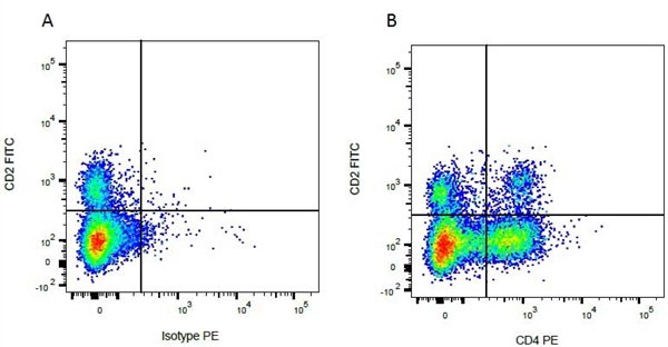
ARG23427 anti-CD4 antibody [CC8] (PE) FACS image
Flow Cytometry: Figure A. FITC conjugated Mouse anti Bovine CD2 and PE-conjugated Mouse IgG2a isotype control. Figure B. FITC conjugated Mouse anti Bovine CD2 and ARG23427 anti-CD4 antibody [CC8] (PE). All experiments performed on red cell lysed Bovine blood gated on mononuclear cells.
-
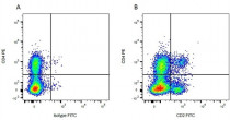
ARG23427 anti-CD4 antibody [CC8] (PE) FACS image
Flow Cytometry: Figure A. ARG23427 anti-CD4 antibody [CC8] (PE) and FITC conjugated Mouse IgG1 isotype control. Figure B. ARG23427 anti-CD4 antibody [CC8] (PE) and FITC conjugated Mouse anti Bovine CD2. All experiments performed on red cell lysed Bovine blood gated on mononuclear cells.
-

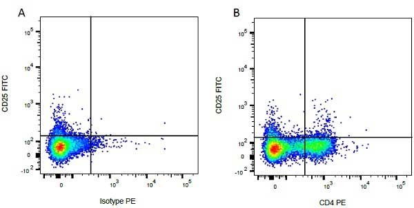
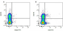
ARG23427 anti-CD4 antibody [CC8] (PE) FACS image
Flow Cytometry: Figure A. FITC conjugated Mouse anti Bovine CD25 and PE-conjugated Mouse IgG2a isotype control. Figure B. FITC conjugated Mouse anti Bovine CD25 and ARG23427 anti-CD4 antibody [CC8] (PE). All experiments performed on red cell lysed Bovine blood gated on mononuclear cells.
-
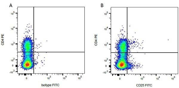
ARG23427 anti-CD4 antibody [CC8] (PE) FACS image
Flow Cytometry: Figure A. ARG23427 anti-CD4 antibody [CC8] (PE) and FITC conjugated Mouse IgG1 isotype control. Figure B. ARG23427 anti-CD4 antibody [CC8] (PE) and FITC conjugated Mouse anti Bovine CD25. All experiments performed on red cell lysed Bovine blood gated on mononuclear cells.