ARG20819
anti-CD3e antibody [C363.29B] (PE)
anti-CD3e antibody [C363.29B] (PE) for Cellular activation ,Depletion,Flow cytometry,IHC-Frozen sections,IHC-Formalin-fixed paraffin-embedded sections and Mouse
Cancer antibody; Developmental Biology antibody; Immune System antibody; Lymphocyte Marker antibody; Inflammatory Cell Marker antibody; T-cell Marker antibody; T-cell infiltration Study antibody; Tumor-infiltrating Lymphocyte Study antibody
Overview
| Product Description | PE-conjugated Rat Monoclonal antibody [C363.29B] recognizes CD3e |
|---|---|
| Tested Reactivity | Ms |
| Tested Application | Cell-Act , Depletion, FACS, IHC-Fr, IHC-P |
| Specificity | Mouse CD3ε. The clone C363.29B recognizes an epitope on the 25 kDa ε chain of the CD3/TCR complex. In the presence of Fc receptor-bearing accessory cells, soluble C363.29B can activate primed and naïve T cell in vitro. Immobilized C363.2B9 monoclonal antibody can also activate both normal T lymphocytes and cloned T cell lines provided the appropriate accessory signals are present. |
| Host | Rat |
| Clonality | Monoclonal |
| Clone | C363.29B |
| Isotype | IgG2b, kappa |
| Target Name | CD3e |
| Antigen Species | Mouse |
| Immunogen | IL-4 producing Th2 cell lines including D10 |
| Conjugation | PE |
| Alternate Names | T-cell surface antigen T3/Leu-4 epsilon chain; T3E; TCRE; T-cell surface glycoprotein CD3 epsilon chain; IMD18; CD antigen CD3e |
Application Instructions
| Application Suggestion |
|
||||||||||||
|---|---|---|---|---|---|---|---|---|---|---|---|---|---|
| Application Note | * The dilutions indicate recommended starting dilutions and the optimal dilutions or concentrations should be determined by the scientist. |
Properties
| Form | Liquid |
|---|---|
| Buffer | PBS, 0.1% Sodium azide and Sucrose. |
| Preservative | 0.1% Sodium azide |
| Stabilizer | Sucrose |
| Concentration | 0.1 mg/ml |
| Storage Instruction | Aliquot and store in the dark at 2-8°C. Keep protected from prolonged exposure to light. Avoid repeated freeze/thaw cycles. Suggest spin the vial prior to opening. The antibody solution should be gently mixed before use. |
| Note | For laboratory research only, not for drug, diagnostic or other use. |
Bioinformation
| Database Links |
Swiss-port # P22646 Mouse T-cell surface glycoprotein CD3 epsilon chain |
|---|---|
| Gene Symbol | CD3E |
| Gene Full Name | CD3 antigen, epsilon polypeptide |
| Background | CD3 subunit complex is crucial in transducing antigen-recognition signals into the cytoplasm of T cells and in regulating the cell surface expression of the TCR complex. T cell activation through the antigen receptor (TCR) involves the cytoplasmic tails of the CD3 subunits CD3 gamma, CD3 delta, CD3 epsilon and CD3 zeta. These CD3 subunits are structurally related members of the immunoglobulins superfamily encoded by closely linked genes on human chromosome 11. The CD3 components have long cytoplasmic tails that associate with cytoplasmic signal transduction molecules. This association is mediated at least in part by a double tyrosine-based motif present in a single copy in the CD3 subunits. CD3 may play a role in TCR-induced growth arrest, cell survival and proliferation. |
| Function | CD3: Part of the TCR-CD3 complex present on T-lymphocyte cell surface that plays an essential role in adaptive immune response. When antigen presenting cells (APCs) activate T-cell receptor (TCR), TCR-mediated signals are transmitted across the cell membrane by the CD3 chains CD3D, CD3E, CD3G and CD3Z. All CD3 chains contain immunoreceptor tyrosine-based activation motifs (ITAMs) in their cytoplasmic domain. Upon TCR engagement, these motifs become phosphorylated by Src family protein tyrosine kinases LCK and FYN, resulting in the activation of downstream signaling pathways (PubMed:2470098). In addition of this role of signal transduction in T-cell activation, CD3E plays an essential role in correct T-cell development. Initiates the TCR-CD3 complex assembly by forming the two heterodimers CD3D/CD3E and CD3G/CD3E. Participates also in internalization and cell surface down-regulation of TCR-CD3 complexes via endocytosis sequences present in CD3E cytosolic region (PubMed:10384095, PubMed:26507128). [UniProt] |
| Highlight | Related products: CD3 antibodies; CD3 ELISA Kits; CD3 Duos / Panels; Anti-Rat IgG secondary antibodies; Related news: New antibody panels and duos for Tumor immune microenvironment Tumor-Infiltrating Lymphocytes (TILs) |
| Research Area | Cancer antibody; Developmental Biology antibody; Immune System antibody; Lymphocyte Marker antibody; Inflammatory Cell Marker antibody; T-cell Marker antibody; T-cell infiltration Study antibody; Tumor-infiltrating Lymphocyte Study antibody |
| Calculated MW | 23 kDa |
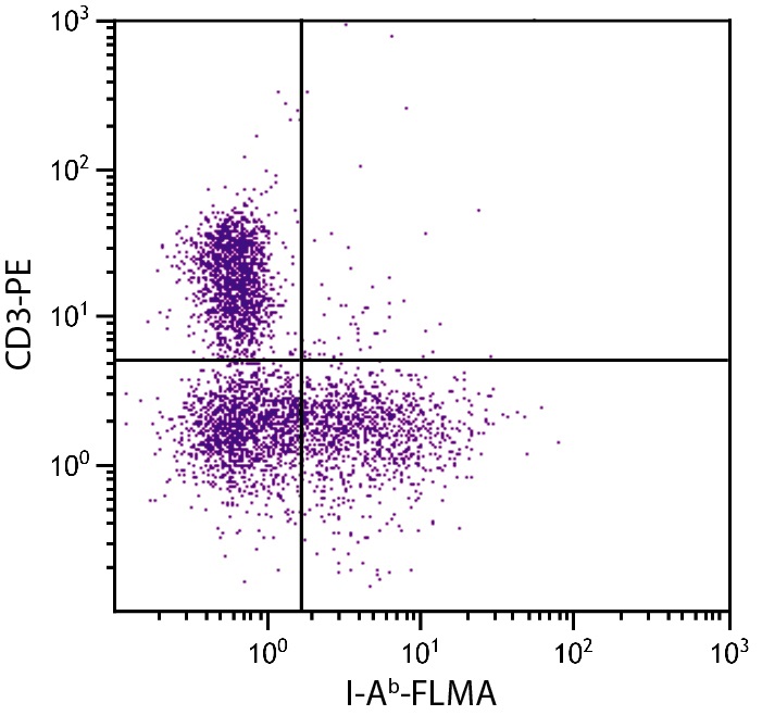
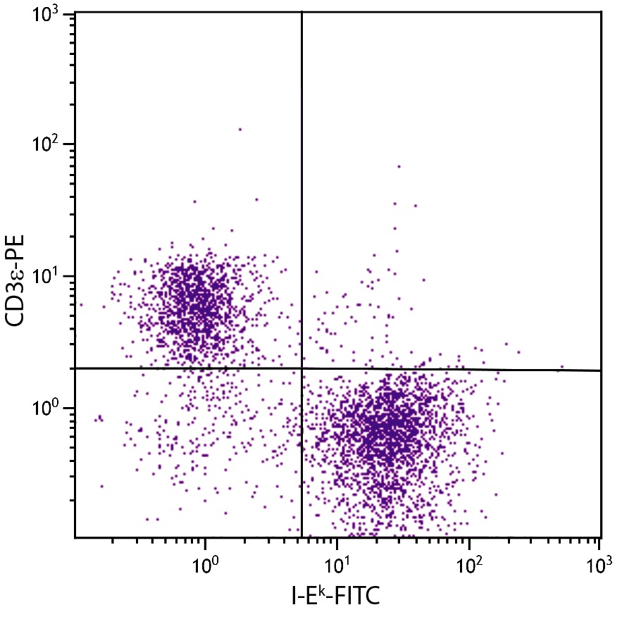
Images (15) Click the Picture to Zoom In
-

-
-
-
-
-
-
-
-
-
-
-

-
-
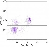
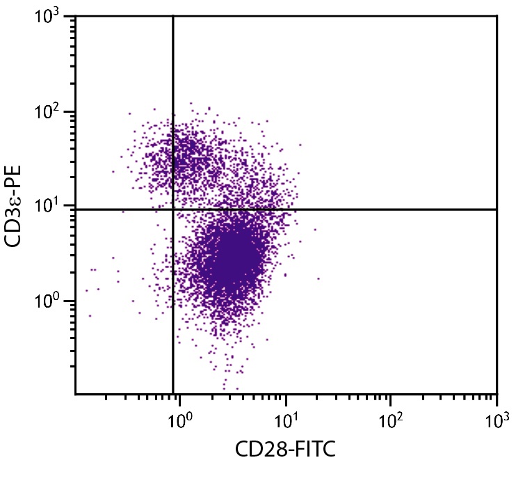
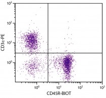
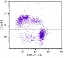
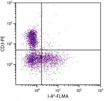
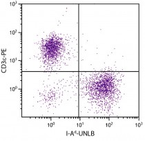
ARG20819 anti-CD3e antibody [C363.29B] (PE) FACS image
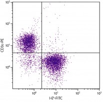
Flow Cytometry: BALB/c Mouse splenocytes stained with ARG22333 anti-IL2 Receptor beta antibody [5H4] (FITC) and ARG20819 anti-CD3e antibody [C363.29B] (PE).
-
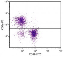
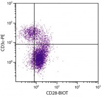
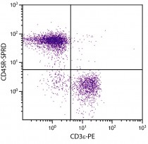
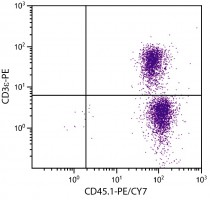
ARG20819 anti-CD3e antibody [C363.29B] (PE) FACS image
Flow Cytometry: BALB/c Mouse splenocytes stained with ARG20819 anti-CD3e antibody [C363.29B] (PE) and ARG20850 anti-CD19 antibody [6D5] (FITC).