ARG54744
anti-CD3 epsilon antibody [145-2C11] (low endotoxin)
anti-CD3 epsilon antibody [145-2C11] (low endotoxin) for Blocking,Cellular activation ,Depletion,Flow cytometry,ICC/IF,IHC-Frozen sections,Immunoprecipitation and Mouse
Cancer antibody; Developmental Biology antibody; Immune System antibody; Lymphocyte Marker antibody; Inflammatory Cell Marker antibody; T-cell Marker antibody; T-cell infiltration Study antibody; Tumor-infiltrating Lymphocyte Study antibody
Overview
| Product Description | Low endotoxin Hamster Monoclonal antibody [145-2C11] recognizes CD3 epsilon |
|---|---|
| Tested Reactivity | Ms |
| Tested Application | BL, Cell-Act , Depletion, FACS, ICC/IF, IHC-Fr, IP |
| Specificity | The Armenian hamster monoclonal antibody 145-2C11 reacts with mouse CD3 (epsilon subunit). This antibody is commonly used as a phenotypic marker for mouse T cells. |
| Host | Hamster |
| Clonality | Monoclonal |
| Clone | 145-2C11 |
| Isotype | IgG |
| Target Name | CD3 epsilon |
| Antigen Species | Mouse |
| Immunogen | Mouse BM10-37 cytotoxic T lymphocytes. |
| Conjugation | Un-conjugated |
| Alternate Names | T-cell surface antigen T3/Leu-4 epsilon chain; T3E; TCRE; T-cell surface glycoprotein CD3 epsilon chain; IMD18; CD antigen CD3e |
Application Instructions
| Application Suggestion |
|
||||||||||||||||
|---|---|---|---|---|---|---|---|---|---|---|---|---|---|---|---|---|---|
| Application Note | Functional Application: Induction of T cell activation, proliferation or apoptosis (depending on conditions); in vivo T cell depletion. * The dilutions indicate recommended starting dilutions and the optimal dilutions or concentrations should be determined by the scientist. |
Properties
| Form | Liquid |
|---|---|
| Purification | Purification with Protein G. |
| Purification Note | 0.2 µm filter sterilized. Endotoxin level is <0.01 EU/µg of the protein, as determined by the LAL test. |
| Purity | > 95% (by SDS-PAGE) |
| Buffer | PBS (pH 7.4) |
| Concentration | 1 mg/ml |
| Storage Instruction | For continuous use, store undiluted antibody at 2-8°C for up to a week. For long-term storage, aliquot and store at -20°C or below. Storage in frost free freezers is not recommended. Avoid repeated freeze/thaw cycles. Suggest spin the vial prior to opening. The antibody solution should be gently mixed before use. |
| Note | For laboratory research only, not for drug, diagnostic or other use. |
Bioinformation
| Database Links |
Swiss-port # P22646 Mouse T-cell surface glycoprotein CD3 epsilon chain |
|---|---|
| Gene Symbol | Cd3e |
| Gene Full Name | CD3 antigen, epsilon polypeptide |
| Background | CD3 subunit complex is crucial in transducing antigen-recognition signals into the cytoplasm of T cells and in regulating the cell surface expression of the TCR complex. T cell activation through the antigen receptor (TCR) involves the cytoplasmic tails of the CD3 subunits CD3 gamma, CD3 delta, CD3 epsilon and CD3 zeta. These CD3 subunits are structurally related members of the immunoglobulins superfamily encoded by closely linked genes on human chromosome 11. The CD3 components have long cytoplasmic tails that associate with cytoplasmic signal transduction molecules. This association is mediated at least in part by a double tyrosine-based motif present in a single copy in the CD3 subunits. CD3 may play a role in TCR-induced growth arrest, cell survival and proliferation. |
| Function | CD3: Part of the TCR-CD3 complex present on T-lymphocyte cell surface that plays an essential role in adaptive immune response. When antigen presenting cells (APCs) activate T-cell receptor (TCR), TCR-mediated signals are transmitted across the cell membrane by the CD3 chains CD3D, CD3E, CD3G and CD3Z. All CD3 chains contain immunoreceptor tyrosine-based activation motifs (ITAMs) in their cytoplasmic domain. Upon TCR engagement, these motifs become phosphorylated by Src family protein tyrosine kinases LCK and FYN, resulting in the activation of downstream signaling pathways (PubMed:2470098). In addition of this role of signal transduction in T-cell activation, CD3E plays an essential role in correct T-cell development. Initiates the TCR-CD3 complex assembly by forming the two heterodimers CD3D/CD3E and CD3G/CD3E. Participates also in internalization and cell surface down-regulation of TCR-CD3 complexes via endocytosis sequences present in CD3E cytosolic region (PubMed:10384095, PubMed:26507128). [UniProt] |
| Highlight | Related products: CD3 antibodies; CD3 ELISA Kits; CD3 Duos / Panels; CD3 recombinant proteins; Anti-Hamster IgG secondary antibodies; Related news: New antibody panels and duos for Tumor immune microenvironment Tumor-Infiltrating Lymphocytes (TILs) Exploring Antiviral Immune Response |
| Research Area | Cancer antibody; Developmental Biology antibody; Immune System antibody; Lymphocyte Marker antibody; Inflammatory Cell Marker antibody; T-cell Marker antibody; T-cell infiltration Study antibody; Tumor-infiltrating Lymphocyte Study antibody |
| Calculated MW | 23 kDa |
Images (2) Click the Picture to Zoom In
-
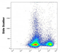
ARG54744 anti-CD3 epsilon antibody [145-2C11] (low endotoxin) FACS image
Flow Cytometry: Murine splenocyte suspension stained with ARG54744 anti-CD3 epsilon antibody [145-2C11] (low endotoxin) at 4 µg/ml dilution, followed by APC-conjugated Donkey anti-Rat antibody.
-
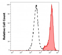
ARG54744 anti-CD3 epsilon antibody [145-2C11] (low endotoxin) FACS image
Flow Cytometry: Separation of murine CD3 positive splenocytes (red-filled) from CD3 negative splenocytes (black-dashed). Murine splenocyte suspension stained with ARG54744 anti-CD3 epsilon antibody [145-2C11] (low endotoxin) at 4 µg/ml dilution, followed by APC-conjugated Donkey anti-Rat antibody.


Clone References
Statins induce regulatory T cell recruitment via a CCL1 dependent pathway.
IHC-Fr / Mouse


HSP70 vaccine in combination with gene therapy with plasmid DNA encoding sPD-1 overcomes immune resistance and suppresses the progression of pulmonary metastatic melanoma.
Functional analysis of B and T lymphocyte attenuator engagement on CD4+ and CD8+ T cells.
CCR5-deficient mice develop experimental autoimmune uveoretinitis in the context of a deviant effector response.
Regulation of NK cell function in vivo by the dose of tumour transplanted in the peritoneum.
FACS / Rat


