ARG53823
anti-CD3 epsilon (activation epitope) antibody [APA1/1] (PE)
anti-CD3 epsilon (activation epitope) antibody [APA1/1] (PE) for Flow cytometry and Human,Mouse
Cancer antibody; Developmental Biology antibody; Immune System antibody; Lymphocyte Marker antibody; Inflammatory Cell Marker antibody; T-cell Marker antibody; T-cell infiltration Study antibody; Tumor-infiltrating Lymphocyte Study antibody
Overview
| Product Description | PE-conjugated Mouse Monoclonal antibody [APA1/1] recognizes CD3 epsilon (activation epitope) |
|---|---|
| Tested Reactivity | Hu, Ms |
| Tested Application | FACS |
| Specificity | The clone APA1/1 recognizes an activation-dependent intracellular epitope of CD3 epsilon. Exposure of the epitope precedes CD3 phosphorylation and recruitment and activation of ZAP70, which initiates the signaling cascade produced by T-cell activation. APA1/1 provides the earliest known marker for TCR-mediated T cell activation. |
| Host | Mouse |
| Clonality | Monoclonal |
| Clone | APA1/1 |
| Isotype | IgG1 |
| Target Name | CD3 epsilon (activation epitope) |
| Antigen Species | Human |
| Immunogen | Purified human CD3 epsilon proteins isolated from thymus. |
| Conjugation | PE |
| Alternate Names | CD3E; CD3 Epsilon Subunit Of T-Cell Receptor Complex; T-Cell Surface Glycoprotein CD3 Epsilon Chain; CD3e Antigen, Epsilon Polypeptide (TiT3 Complex); T-Cell Surface Antigen T3/Leu-4 Epsilon Chain; CD3e Molecule, Epsilon (CD3-TCR Complex); CD3-Epsilon; CD3epsilon |
Application Instructions
| Application Suggestion |
|
||||
|---|---|---|---|---|---|
| Application Note | * The dilutions indicate recommended starting dilutions and the optimal dilutions or concentrations should be determined by the scientist. |
Properties
| Form | Liquid |
|---|---|
| Purification Note | The purified antibody is conjugated with R-Phycoerythrin (PE) under optimum conditions. The conjugate is purified by size-exclusion chromatography. |
| Buffer | PBS, 15 mM Sodium azide and 0.2% (w/v) high-grade protease free BSA |
| Preservative | 15 mM Sodium azide |
| Stabilizer | 0.2% (w/v) high-grade protease free BSA |
| Concentration | 0.1 mg/ml |
| Storage Instruction | Aliquot and store in the dark at 2-8°C. Keep protected from prolonged exposure to light. Avoid repeated freeze/thaw cycles. Suggest spin the vial prior to opening. The antibody solution should be gently mixed before use. |
| Note | For laboratory research only, not for drug, diagnostic or other use. |
Bioinformation
| Database Links |
Swiss-port # P07766 Human T-cell surface glycoprotein CD3 epsilon chain Swiss-port # P22646 Mouse T-cell surface glycoprotein CD3 epsilon chain |
|---|---|
| Gene Symbol | CD3E |
| Gene Full Name | CD3 Epsilon Subunit Of T-Cell Receptor Complex |
| Background | The protein encoded by this gene is the CD3-epsilon polypeptide, which together with CD3-gamma, -delta and -zeta, and the T-cell receptor alpha/beta and gamma/delta heterodimers, forms the T-cell receptor-CD3 complex. This complex plays an important role in coupling antigen recognition to several intracellular signal-transduction pathways. The genes encoding the epsilon, gamma and delta polypeptides are located in the same cluster on chromosome 11. The epsilon polypeptide plays an essential role in T-cell development. Defects in this gene cause immunodeficiency. This gene has also been linked to a susceptibility to type I diabetes in women. |
| Function | Part of the TCR-CD3 complex present on T-lymphocyte cell surface that plays an essential role in adaptive immune response. When antigen presenting cells (APCs) activate T-cell receptor (TCR), TCR-mediated signals are transmitted across the cell membrane by the CD3 chains CD3D, CD3E, CD3G and CD3Z. All CD3 chains contain immunoreceptor tyrosine-based activation motifs (ITAMs) in their cytoplasmic domain. Upon TCR engagement, these motifs become phosphorylated by Src family protein tyrosine kinases LCK and FYN, resulting in the activation of downstream signaling pathways. |
| Cellular Localization | Cell membrane, Membrane |
| Highlight | Related products: CD3 antibodies; CD3 ELISA Kits; CD3 Duos / Panels; Anti-Mouse IgG secondary antibodies; Related news: New antibody panels and duos for Tumor immune microenvironment Tumor-Infiltrating Lymphocytes (TILs) |
| Research Area | Cancer antibody; Developmental Biology antibody; Immune System antibody; Lymphocyte Marker antibody; Inflammatory Cell Marker antibody; T-cell Marker antibody; T-cell infiltration Study antibody; Tumor-infiltrating Lymphocyte Study antibody |
| Calculated MW | 19 kDa |
Images (1) Click the Picture to Zoom In
-
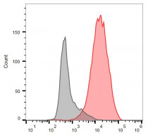
ARG53823 anti-CD3 epsilon (activation epitope) antibody [APA1/1] (PE) FACS image
Flow Cytometry: Separation of stained Jurkat cells (red) from unstained Jurkat cells (grey). Cells were stained with ARG53823 anti-CD3 epsilon (activation epitope) antibody [APA1/1] (PE) at 3 µg/ml dilution.


