ARG55554
anti-CD206 / MMR antibody [15-2]
anti-CD206 / MMR antibody [15-2] for CyTOF®-candidate,Flow cytometry,Functional study,ICC/IF,IHC-Frozen sections,Immunoprecipitation,Western blot and Human,Mouse
Immune System antibody; M1/M2/TAM Marker antibody; Macrophage Marker antibody; M2 Macrophage Marker antibody
Overview
| Product Description | Mouse Monoclonal antibody [15-2] recognizes CD206 / MMR |
|---|---|
| Tested Reactivity | Hu, Ms |
| Tested Application | CyTOF®-candidate, FACS, FuncSt, ICC/IF, IHC-Fr, IP, WB |
| Specificity | This antibody recognizes an extracellular epitope of CD206 (macrophage mannose receptor, MMR), a 162-175 kDa type I transmembrane protein expressed mainly on macrophages, dendritic cells and hepatic or lymphatic endothelial cells, but not on monocytes. |
| Host | Mouse |
| Clonality | Monoclonal |
| Clone | 15-2 |
| Isotype | IgG1 |
| Target Name | CD206 / MMR |
| Antigen Species | Human |
| Immunogen | Purified Human mannose receptor (NP_002429.1) |
| Conjugation | Un-conjugated |
| Alternate Names | CLEC13D; C-type lectin domain family 13 member D; Macrophage mannose receptor 1-like protein 1; C-type lectin domain family 13 member D-like; MMR; CLEC13DL; CD206; Macrophage mannose receptor 1; bA541I19.1; CD antigen CD206; MRC1L1 |
Application Instructions
| Application Suggestion |
|
||||||||||||||||
|---|---|---|---|---|---|---|---|---|---|---|---|---|---|---|---|---|---|
| Application Note | IHC-Fr: Incubate with the antibody at 4°C for overnight. * The dilutions indicate recommended starting dilutions and the optimal dilutions or concentrations should be determined by the scientist. |
Properties
| Form | Liquid |
|---|---|
| Purification | Purification with Protein A. |
| Purity | > 95% (by SDS-PAGE) |
| Buffer | PBS (pH 7.4) and 15 mM Sodium azide |
| Preservative | 15 mM Sodium azide |
| Concentration | 1 mg/ml |
| Storage Instruction | For continuous use, store undiluted antibody at 2-8°C for up to a week. For long-term storage, aliquot and store at -20°C or below. Storage in frost free freezers is not recommended. Avoid repeated freeze/thaw cycles. Suggest spin the vial prior to opening. The antibody solution should be gently mixed before use. |
| Note | For laboratory research only, not for drug, diagnostic or other use. |
Bioinformation
| Database Links | |
|---|---|
| Gene Symbol | MRC1 |
| Gene Full Name | mannose receptor, C type 1 |
| Background | The recognition of complex carbohydrate structures on glycoproteins is an important part of several biological processes, including cell-cell recognition, serum glycoprotein turnover, and neutralization of pathogens. CD206 / MMR is a type I membrane receptor that mediates the endocytosis of glycoproteins by macrophages. The protein has been shown to bind high-mannose structures on the surface of potentially pathogenic viruses, bacteria, and fungi so that they can be neutralized by phagocytic engulfment. [provided by RefSeq, Sep 2015] |
| Function | CD206 / MMR mediates the endocytosis of glycoproteins by macrophages. Binds both sulfated and non-sulfated polysaccharide chains. (Microbial infection) Acts as phagocytic receptor for bacteria, fungi and other pathogens. (Microbial infection) Acts as a receptor for Dengue virus envelope protein E. (Microbial infection) Interacts with Hepatitis B virus envelope protein. [UniProt] |
| Highlight | Related Antibody Duos and Panels: ARG30333 M1/M2/TAM Marker Antibody Panel Related products: CD206 antibodies; CD206 ELISA Kits; CD206 Duos / Panels; Anti-Mouse IgG secondary antibodies; Related news: CyTOF-candidate Antibodies New antibody panels and duos for Tumor immune microenvironment Tumor-Infiltrating Lymphocytes (TILs) Anti-SerpinB9 therapy, a new strategy for cancer therapy RIP1 activation and pathogenesis of NASH |
| Research Area | Immune System antibody; M1/M2/TAM Marker antibody; Macrophage Marker antibody; M2 Macrophage Marker antibody |
| Calculated MW | 166 kDa |
Images (3) Click the Picture to Zoom In
-
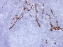
ARG55554 anti-CD206 / MMR antibody [15-2] IHC-Fr image
Immunohistochemistry: Frozen section of Human tonsil tissue stained with ARG55554 anti-CD206 / MMR antibody [15-2] at 1:25 dilution. The antibody stains endothelia of lymph vessels strongly.
-
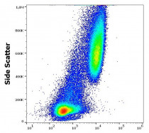
ARG55554 anti-CD206 / MMR antibody [15-2] FACS image
Flow Cytometry: Stimulated (GM-CSF + IL-4) human peripheral blood mononuclear cells stained with ARG55554 anti-CD206 / MMR antibody [15-2] at 9 µg/ml dilution, followed by PE-conjugated Goat anti-Mouse antibody.
-
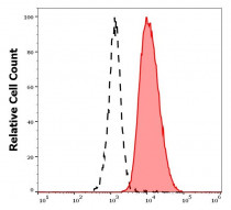
ARG55554 anti-CD206 / MMR antibody [15-2] FACS image
Flow Cytometry: Separation of human CD206 positive dendritic cells differentiated upon monocyte stimulation (GM-CSF + IL-4) (red-filled) from non-stimulated lymphocytes (black-dashed). Human stimulated (GM-CSF + IL-4) peripheral blood mononuclear cells stained with ARG55554 anti-CD206 / MMR antibody [15-2] at 9 µg/ml dilution, followed by PE-conjugated Goat anti-Mouse antibody.


Specific References
Dual keratinocyte-attachment and anti-inflammatory coatings for soft tissue sealing around transmucosal oral implants
ICC/IF / Mouse


