ARG41947
anti-CD206 / MMR antibody [15-2] (APC)
anti-CD206 / MMR antibody [15-2] (APC) for Flow cytometry and Human
Immune System antibody; M1/M2/TAM Marker antibody; Macrophage Marker antibody; M2 Macrophage Marker antibody
Overview
| Product Description | APC-conjugated Mouse Monoclonal antibody [15-2] recognizes CD206 / MMR |
|---|---|
| Tested Reactivity | Hu |
| Tested Application | FACS |
| Host | Mouse |
| Clonality | Monoclonal |
| Clone | 15-2 |
| Isotype | IgG1, kappa |
| Target Name | CD206 / MMR |
| Antigen Species | Human |
| Immunogen | Purified Human CD206 / MMR. |
| Conjugation | APC |
| Alternate Names | CLEC13D; C-type lectin domain family 13 member D; Macrophage mannose receptor 1-like protein 1; C-type lectin domain family 13 member D-like; MMR; CLEC13DL; CD206; Macrophage mannose receptor 1; bA541I19.1; CD antigen CD206; MRC1L1 |
Application Instructions
| Application Suggestion |
|
||||
|---|---|---|---|---|---|
| Application Note | * The dilutions indicate recommended starting dilutions and the optimal dilutions or concentrations should be determined by the scientist. |
Properties
| Form | Liquid |
|---|---|
| Purification | Purified |
| Buffer | PBS and 15 mM Sodium azide. |
| Preservative | 15 mM Sodium azide |
| Storage Instruction | Aliquot and store in the dark at 2-8°C. Keep protected from prolonged exposure to light. Avoid repeated freeze/thaw cycles. Suggest spin the vial prior to opening. The antibody solution should be gently mixed before use. |
| Note | For laboratory research only, not for drug, diagnostic or other use. |
Bioinformation
| Database Links | |
|---|---|
| Gene Symbol | MRC1 |
| Gene Full Name | mannose receptor, C type 1 |
| Background | The recognition of complex carbohydrate structures on glycoproteins is an important part of several biological processes, including cell-cell recognition, serum glycoprotein turnover, and neutralization of pathogens. CD206 / MMR is a type I membrane receptor that mediates the endocytosis of glycoproteins by macrophages. The protein has been shown to bind high-mannose structures on the surface of potentially pathogenic viruses, bacteria, and fungi so that they can be neutralized by phagocytic engulfment. [provided by RefSeq, Sep 2015] |
| Function | CD206 / MMR mediates the endocytosis of glycoproteins by macrophages. Binds both sulfated and non-sulfated polysaccharide chains. (Microbial infection) Acts as phagocytic receptor for bacteria, fungi and other pathogens. (Microbial infection) Acts as a receptor for Dengue virus envelope protein E. (Microbial infection) Interacts with Hepatitis B virus envelope protein. [UniProt] |
| Cellular Localization | Endosome membrane; Single-pass type I membrane protein. Cell membrane; Single-pass type I membrane protein. [UniProt] |
| Highlight | Related products: CD206 antibodies; CD206 ELISA Kits; CD206 Duos / Panels; Anti-Mouse IgG secondary antibodies; Related news: New antibody panels and duos for Tumor immune microenvironment Tumor-Infiltrating Lymphocytes (TILs) Anti-SerpinB9 therapy, a new strategy for cancer therapy RIP1 activation and pathogenesis of NASH |
| Research Area | Immune System antibody; M1/M2/TAM Marker antibody; Macrophage Marker antibody; M2 Macrophage Marker antibody |
| Calculated MW | 166 kDa |
Images (3) Click the Picture to Zoom In
-
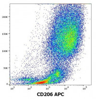
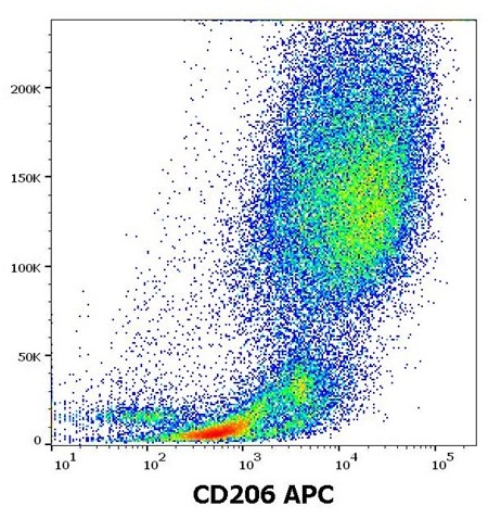
ARG41947 anti-CD206 / MMR antibody [15-2] (APC) FACS image
Flow Cytometry: Stimulated Human monocytes (GM-CSF + IL4) stained with ARG41947 anti-CD206 / MMR antibody [15-2] (APC) at 10 µl / 10^6 cells in 100 µl of cell suspension.
-
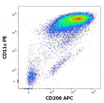
ARG41947 anti-CD206 / MMR antibody [15-2] (APC) FACS image
Flow Cytometry: Stimulated Human monocytes (GM-CSF + IL4) stained with ARG41947 anti-CD206 / MMR antibody [15-2] (APC) at 10 µl / 10^6 cells in 100 µl of cell suspension and ARG53762 anti-CD11c antibody [BU15] (PE) at 20 µl / 10^6 cells in 100 µl of cell suspension.
-
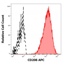
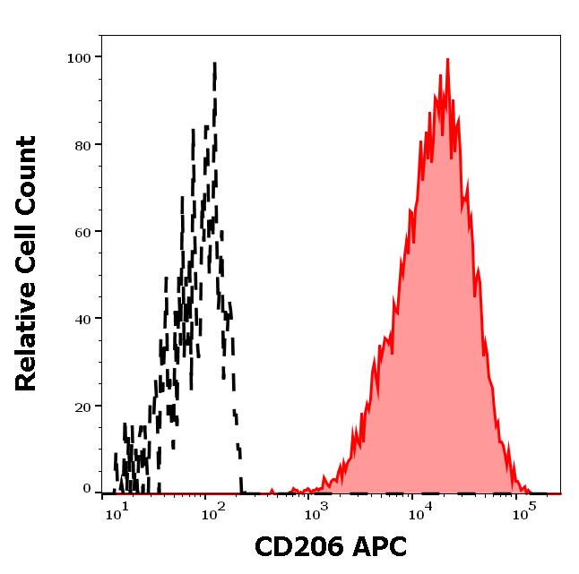
ARG41947 anti-CD206 / MMR antibody [15-2] (APC) FACS image
Flow Cytometry: Separation of Human CD206 positive CD11c positive dendritic cells differentiated upon monocyte stimulation (GM-CSF + IL4) (red-filled) from non-stimulated lymphocytes (black-dashed). Cells were stained with ARG41947 anti-CD206 / MMR antibody [15-2] (APC) at 10 µl / 100 µl of peripheral whole blood.