ARG62766
anti-CD20 antibody [LT20] (Biotin)
anti-CD20 antibody [LT20] (Biotin) for Flow cytometry and Human
Cancer antibody; Developmental Biology antibody; Immune System antibody; B cell Marker antibody; Immature B Cell Marker antibody; Inflammatory Cell Marker antibody; Tumor-infiltrating Lymphocyte Study antibody
Overview
| Product Description | Biotin-conjugated Mouse Monoclonal antibody [LT20] recognizes CD20 |
|---|---|
| Tested Reactivity | Hu |
| Tested Application | FACS |
| Specificity | The clone LT20 reacts with CD20 (Bp35), a 33-37 kDa non-glycosylated membrane receptor with four transmembrane domains, expressed on B lymphocytes (it is lost on plasma cells), follicular dendritic cells, and at low levels on peripheral blood T lymphocytes. |
| Host | Mouse |
| Clonality | Monoclonal |
| Clone | LT20 |
| Isotype | IgG2a |
| Target Name | CD20 |
| Antigen Species | Human |
| Immunogen | Normal human lymphocytes from lymph node. |
| Conjugation | Biotin |
| Alternate Names | Bp35; LEU-16; B-lymphocyte surface antigen B1; B-lymphocyte antigen CD20; CD20; S7; CD antigen CD20; Leukocyte surface antigen Leu-16; B1; CVID5; Membrane-spanning 4-domains subfamily A member 1; MS4A2 |
Application Instructions
| Application Suggestion |
|
||||
|---|---|---|---|---|---|
| Application Note | * The dilutions indicate recommended starting dilutions and the optimal dilutions or concentrations should be determined by the scientist. |
Properties
| Form | Liquid |
|---|---|
| Purification Note | The purified antibody is conjugated with Biotin-LC-NHS under optimum conditions. The reagent is free of unconjugated biotin. |
| Buffer | PBS (pH 7.4) and 15 mM Sodium azide |
| Preservative | 15 mM Sodium azide |
| Concentration | 1 mg/ml |
| Storage Instruction | Aliquot and store in the dark at 2-8°C. Keep protected from prolonged exposure to light. Avoid repeated freeze/thaw cycles. Suggest spin the vial prior to opening. The antibody solution should be gently mixed before use. |
| Note | For laboratory research only, not for drug, diagnostic or other use. |
Bioinformation
| Database Links | |
|---|---|
| Gene Symbol | MS4A1 |
| Gene Full Name | membrane-spanning 4-domains, subfamily A, member 1 |
| Background | CD20 is a member of the membrane-spanning 4A gene family. Members of this nascent protein family are characterized by common structural features and similar intron/exon splice boundaries and display unique expression patterns among hematopoietic cells and nonlymphoid tissues. This gene encodes a B-lymphocyte surface molecule which plays a role in the development and differentiation of B-cells into plasma cells. This family member is localized to 11q12, among a cluster of family members. Alternative splicing of this gene results in two transcript variants which encode the same protein. [provided by RefSeq, Jul 2008] |
| Function | CD20 is a B-lymphocyte-specific membrane protein. It plays a role in the regulation of cellular calcium influx necessary for the development, differentiation, and activation of B-lymphocytes (PubMed:3925015, PubMed:7684739, PubMed:12920111). Functions as a store-operated calcium (SOC) channel component promoting calcium influx after activation by the B-cell receptor/BCR (PubMed:7684739, PubMed:12920111, PubMed:18474602). [UniProt] |
| Highlight | Related products: CD20 antibodies; CD20 ELISA Kits; CD20 Duos / Panels; Anti-Mouse IgG secondary antibodies; Related news: New antibody panels and duos for Tumor immune microenvironment Tumor-Infiltrating Lymphocytes (TILs) Exploring Antiviral Immune Response |
| Research Area | Cancer antibody; Developmental Biology antibody; Immune System antibody; B cell Marker antibody; Immature B Cell Marker antibody; Inflammatory Cell Marker antibody; Tumor-infiltrating Lymphocyte Study antibody |
| Calculated MW | 33 kDa |
| PTM | Phosphorylated. Might be functionally regulated by protein kinase(s). |
Images (1) Click the Picture to Zoom In
-
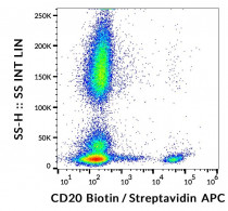
ARG62766 anti-CD20 antibody [LT20] (Biotin) FACS image
Flow Cytometry: Human peripheral blood cells stained with ARG62766 anti-CD20 antibody [LT20] (Biotin), followed by Streptavidin (APC).

Clone References
A differential cell capture assay for evaluating antibody interactions with cell surface targets.


Low numbers of FOXP3 positive regulatory T cells are present in all developmental stages of human atherosclerotic lesions.
IHC / Human
CD20-induced lymphoma cell death is independent of both caspases and its redistribution into triton X-100 insoluble membrane rafts.


