ARG63123
anti-CD20 antibody
anti-CD20 antibody for IHC-Formalin-fixed paraffin-embedded sections,Western blot and Human
Cancer antibody; Developmental Biology antibody; Immune System antibody; B cell Marker antibody; Immature B Cell Marker antibody; Inflammatory Cell Marker antibody; Tumor-infiltrating Lymphocyte Study antibody
Overview
| Product Description | Goat Polyclonal antibody recognizes CD20 |
|---|---|
| Tested Reactivity | Hu |
| Tested Application | IHC-P, WB |
| Specificity | Please note that both reported variants (NP_068769.2 and NP_690605.1) encode the same protein. |
| Host | Goat |
| Clonality | Polyclonal |
| Isotype | IgG |
| Target Name | CD20 |
| Antigen Species | Human |
| Immunogen | CQDQESSPIENDSSP |
| Conjugation | Un-conjugated |
| Alternate Names | Bp35; LEU-16; B-lymphocyte surface antigen B1; B-lymphocyte antigen CD20; CD20; S7; CD antigen CD20; Leukocyte surface antigen Leu-16; B1; CVID5; Membrane-spanning 4-domains subfamily A member 1; MS4A2 |
Application Instructions
| Application Suggestion |
|
||||||
|---|---|---|---|---|---|---|---|
| Application Note | WB: Recommend incubate at RT for 1h. IHC-P: Antigen Retrieval: Steam tissue section in Citrate buffer (pH 6.0). * The dilutions indicate recommended starting dilutions and the optimal dilutions or concentrations should be determined by the scientist. |
Properties
| Form | Liquid |
|---|---|
| Purification | Purified from goat serum by antigen affinity chromatography. |
| Buffer | Tris saline (pH 7.3), 0.02% Sodium azide and 0.5% BSA. |
| Preservative | 0.02% Sodium azide |
| Stabilizer | 0.5% BSA |
| Concentration | 0.5 mg/ml |
| Storage Instruction | For continuous use, store undiluted antibody at 2-8°C for up to a week. For long-term storage, aliquot and store at -20°C or below. Storage in frost free freezers is not recommended. Avoid repeated freeze/thaw cycles. Suggest spin the vial prior to opening. The antibody solution should be gently mixed before use. |
| Note | For laboratory research only, not for drug, diagnostic or other use. |
Bioinformation
| Database Links | |
|---|---|
| Background | CD20 is a member of the membrane-spanning 4A gene family. Members of this nascent protein family are characterized by common structural features and similar intron/exon splice boundaries and display unique expression patterns among hematopoietic cells and nonlymphoid tissues. This gene encodes a B-lymphocyte surface molecule which plays a role in the development and differentiation of B-cells into plasma cells. This family member is localized to 11q12, among a cluster of family members. Alternative splicing of this gene results in two transcript variants which encode the same protein. [provided by RefSeq, Jul 2008] |
| Function | CD20 is a B-lymphocyte-specific membrane protein. It plays a role in the regulation of cellular calcium influx necessary for the development, differentiation, and activation of B-lymphocytes (PubMed:3925015, PubMed:7684739, PubMed:12920111). Functions as a store-operated calcium (SOC) channel component promoting calcium influx after activation by the B-cell receptor/BCR (PubMed:7684739, PubMed:12920111, PubMed:18474602). [UniProt] |
| Highlight | Related products: CD20 antibodies; CD20 ELISA Kits; CD20 Duos / Panels; Anti-Goat IgG secondary antibodies; Related news: New antibody panels and duos for Tumor immune microenvironment Tumor-Infiltrating Lymphocytes (TILs) Exploring Antiviral Immune Response |
| Research Area | Cancer antibody; Developmental Biology antibody; Immune System antibody; B cell Marker antibody; Immature B Cell Marker antibody; Inflammatory Cell Marker antibody; Tumor-infiltrating Lymphocyte Study antibody |
| Calculated MW | 33 kDa |
| PTM | Phosphorylated. Might be functionally regulated by protein kinase(s). |
Images (2) Click the Picture to Zoom In
-
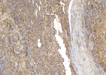
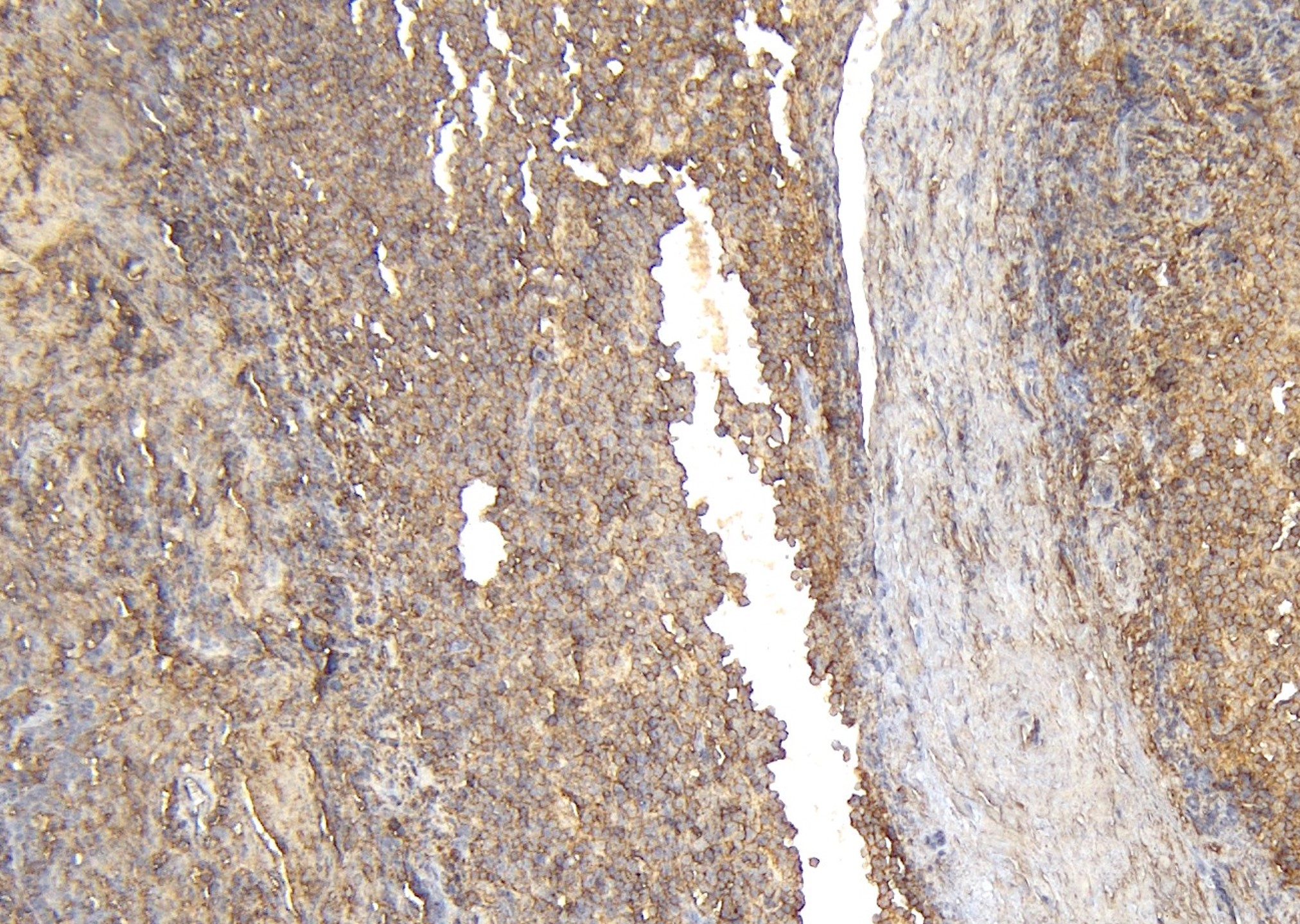
ARG63123 anti-CD20 antibody IHC-P image
Immunohistochemistry: Paraffin-embedded Human tonsil tissue. Antigen Retrieval: Steam tissue section in Citrate buffer (pH 6.0). The tissue section was stained with ARG63123 anti-CD20 antibody at 6 µg/ml.
-
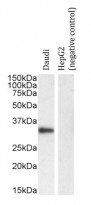
ARG63123 anti-CD20 antibody WB image
Western blot: 35 µg of Daudi and HepG2 (negative control) cell lysates stained with ARG63123 anti-CD20 antibody at 0.01 µg/ml dilution and incubated at RT for 1 hour.