ARG41422
anti-CD18 / LFA1 beta antibody
anti-CD18 / LFA1 beta antibody for Flow cytometry,ICC/IF,IHC-Formalin-fixed paraffin-embedded sections,Western blot and Human
Overview
| Product Description | Rabbit Polyclonal antibody recognizes CD18 / LFA1 beta |
|---|---|
| Tested Reactivity | Hu |
| Tested Application | FACS, ICC/IF, IHC-P, WB |
| Host | Rabbit |
| Clonality | Polyclonal |
| Isotype | IgG |
| Target Name | CD18 / LFA1 beta |
| Antigen Species | Human |
| Immunogen | Recombinant protein corresponding to Q404-S769 of Human CD18. |
| Conjugation | Un-conjugated |
| Alternate Names | MF17; LAD; CD antigen CD18; MFI7; MAC-1; Cell surface adhesion glycoproteins LFA-1/CR3/p150,95 subunit beta; LCAMB; Integrin beta-2; Complement receptor C3 subunit beta; LFA-1; CD18 |
Application Instructions
| Application Suggestion |
|
||||||||||
|---|---|---|---|---|---|---|---|---|---|---|---|
| Application Note | IHC-P: Antigen Retrieval: Heat mediation was performed in Citrate buffer (pH 6.0, epitope retrieval solution) for 20 min. * The dilutions indicate recommended starting dilutions and the optimal dilutions or concentrations should be determined by the scientist. |
||||||||||
| Observed Size | ~ 100 kDa |
Properties
| Form | Liquid |
|---|---|
| Purification | Affinity purification with immunogen. |
| Buffer | 0.2% Na2HPO4, 0.9% NaCl, 0.05% Sodium azide and 5% BSA. |
| Preservative | 0.05% Sodium azide |
| Stabilizer | 5% BSA |
| Concentration | 0.5 mg/ml |
| Storage Instruction | For continuous use, store undiluted antibody at 2-8°C for up to a week. For long-term storage, aliquot and store at -20°C or below. Storage in frost free freezers is not recommended. Avoid repeated freeze/thaw cycles. Suggest spin the vial prior to opening. The antibody solution should be gently mixed before use. |
| Note | For laboratory research only, not for drug, diagnostic or other use. |
Bioinformation
| Database Links | |
|---|---|
| Gene Symbol | ITGB2 |
| Gene Full Name | integrin, beta 2 (complement component 3 receptor 3 and 4 subunit) |
| Background | This gene encodes an integrin beta chain, which combines with multiple different alpha chains to form different integrin heterodimers. Integrins are integral cell-surface proteins that participate in cell adhesion as well as cell-surface mediated signalling. The encoded protein plays an important role in immune response and defects in this gene cause leukocyte adhesion deficiency. Alternative splicing results in multiple transcript variants. [provided by RefSeq, Dec 2014] |
| Function | Integrin alpha-L/beta-2 is a receptor for ICAM1, ICAM2, ICAM3 and ICAM4. Integrins alpha-M/beta-2 and alpha-X/beta-2 are receptors for the iC3b fragment of the third complement component and for fibrinogen. Integrin alpha-X/beta-2 recognizes the sequence G-P-R in fibrinogen alpha-chain. Integrin alpha-M/beta-2 recognizes P1 and P2 peptides of fibrinogen gamma chain. Integrin alpha-M/beta-2 is also a receptor for factor X. Integrin alpha-D/beta-2 is a receptor for ICAM3 and VCAM1. Triggers neutrophil transmigration during lung injury through PTK2B/PYK2-mediated activation. [UniProt] |
| Cellular Localization | Cell membrane; Single-pass type I membrane protein. Membrane raft; Single-pass type I membrane protein. [UniProt] |
| Calculated MW | 85 kDa |
| PTM | Both Ser-745 and Ser-756 become phosphorylated when T-cells are exposed to phorbol esters (PubMed:11700305). Phosphorylation on Thr-758 (but not on Ser-756) allows interaction with 14-3-3 proteins (PubMed:11700305, PubMed:16301335). [UniProt] |
Images (7) Click the Picture to Zoom In
-
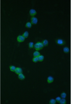
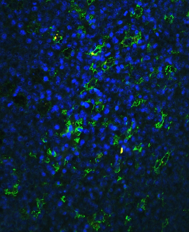
ARG41422 anti-CD18 / LFA1 beta antibody ICC/IF image
Immunofluorescence: THP-1 cells stained with ARG41422 anti-CD18 / LFA1 beta antibody (green) at 2 µg/ml dilution, overnight at 4°C. DAPI (blue) for nuclear staining.
-
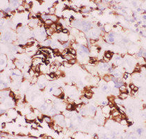
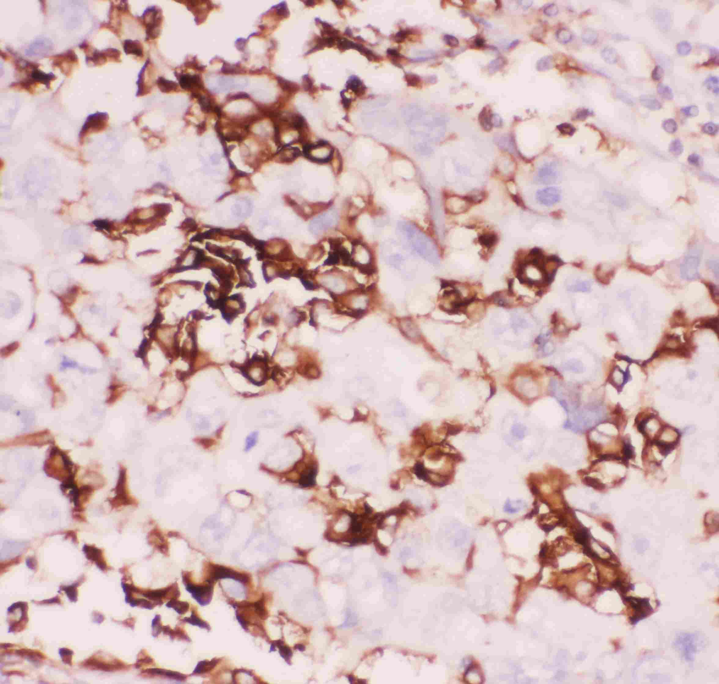
ARG41422 anti-CD18 / LFA1 beta antibody IHC-P image
Immunohistochemistry: Paraffin-embedded Human lung cancer tissue. Antigen Retrieval: Heat mediation was performed in Citrate buffer (pH 6.0, epitope retrieval solution) for 20 min. The tissue section was blocked with 10% goat serum. The tissue section was then stained with ARG41422 anti-CD18 / LFA1 beta antibody at 1 µg/ml dilution, overnight at 4°C.
-
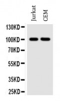
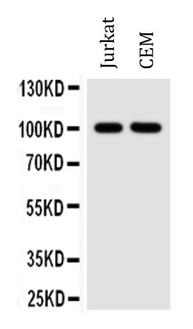
ARG41422 anti-CD18 / LFA1 beta antibody WB image
Western blot: 50 µg of samples under reducing conditions. Jurkat and CEM whole cell lysates stained with ARG41422 anti-CD18 / LFA1 beta antibody at 0.5 µg/ml dilution, overnight at 4°C.
-
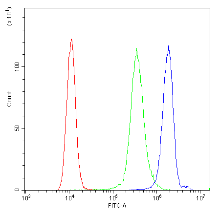
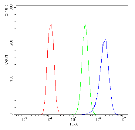
ARG41422 anti-CD18 / LFA1 beta antibody FACS image
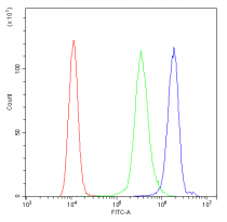
Flow Cytometry: Raji cells were blocked with 10% normal goat serum and then stained with ARG41422 anti-CD18 / LFA1 beta antibody (blue) at 1 µg/10^6 cells for 30 min at 20°C, followed by incubation with DyLight®488 labelled secondary antibody. Isotype control antibody (green) was Rabbit IgG (1 µg/10^6 cells) used under the same conditions. Unlabelled sample (red) was also used as a control.
-

ARG41422 anti-CD18 / LFA1 beta antibody IHC-P image
Immunohistochemistry: Paraffin-embedded Human tonsil tissue. Antigen Retrieval: Heat mediation was performed in Citrate buffer (pH 6.0, epitope retrieval solution) for 20 min. The tissue section was blocked with 10% goat serum. The tissue section was then stained with ARG41422 anti-CD18 / LFA1 beta antibody at 1 µg/ml dilution, overnight at 4°C.
-
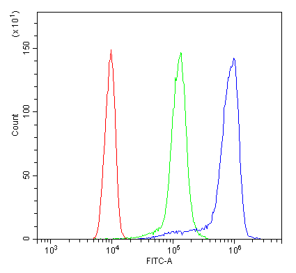
ARG41422 anti-CD18 / LFA1 beta antibody FACS image
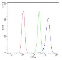
Flow Cytometry: U2OS cells were blocked with 10% normal goat serum and then stained with ARG41422 anti-CD18 / LFA1 beta antibody (blue) at 1 µg/10^6 cells for 30 min at 20°C, followed by incubation with DyLight®488 labelled secondary antibody. Isotype control antibody (green) was Rabbit IgG (1 µg/10^6 cells) used under the same conditions. Unlabelled sample (red) was also used as a control.
-
ARG41422 anti-CD18 / LFA1 beta antibody FACS image
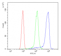
Flow Cytometry: THP-1 cells were blocked with 10% normal goat serum and then stained with ARG41422 anti-CD18 / LFA1 beta antibody (blue) at 1 µg/10^6 cells for 30 min at 20°C, followed by incubation with DyLight®488 labelled secondary antibody. Isotype control antibody (green) was Rabbit IgG (1 µg/10^6 cells) used under the same conditions. Unlabelled sample (red) was also used as a control.