ARG23390
anti-CD163 antibody [ED2]
anti-CD163 antibody [ED2] for Flow cytometry,ICC/IF,IHC-Frozen sections,IHC-Formalin-fixed paraffin-embedded sections,Immunoprecipitation,Western blot and Rat
M1/M2/TAM Marker antibody; Macrophage Marker antibody; M2 Macrophage Marker antibody
Overview
| Product Description | Mouse Monoclonal antibody [ED2] recognizes CD163 Mouse anti Rat CD163, clone ED2 recognises the rat ED2 cell surface glycoprotein (Dijkstra et al. 1985). A 175 kDa molecule also known as rat CD163, a member of the group B scavenger receptor cysteine-rich (SRCR) family and an erythroblast adhesion receptor (Fabriek et al. 2007). Mouse anti rat CD163, clone ED2 was shown to detect approximately 50% of peritoneal macrophages, a subset of splenic macrophages, and most tissue macrophages. However, no staining was observed in monocytes or alveolar macrophages (Dijkstra et al. 1985, Beelen et al. 1987). In freshly isolated bone marrow, expression of CD163 was limited to mature macrophages only (Barbe et al. 1990).Clone ED2 may be used in immunohistology using antigen retrieval, and has also been described reacting with paraffin-embedded material following PLP fixation (Periodate-lysine-paraformaldehyde), see Whiteland et al. |
|---|---|
| Tested Reactivity | Rat |
| Tested Application | FACS, ICC/IF, IHC-Fr, IHC-P, IP, WB |
| Host | Mouse |
| Clonality | Monoclonal |
| Clone | ED2 |
| Isotype | IgG1 |
| Target Name | CD163 |
| Antigen Species | Rat |
| Immunogen | Rat Spleen cell homogenate. |
| Conjugation | Un-conjugated |
| Alternate Names | sCD163; M130; Scavenger receptor cysteine-rich type 1 protein M130; MM130; CD antigen CD163; Hemoglobin scavenger receptor |
Application Instructions
| Application Suggestion |
|
||||||||||||||
|---|---|---|---|---|---|---|---|---|---|---|---|---|---|---|---|
| Application Note | IHC-P: This product requires protein digestion pre-treatment of paraffin sections e.g. trypsin or pronase. FACS: Use 10 µl of the suggested working dilution to label 10^6 cells in 100 µl. * The dilutions indicate recommended starting dilutions and the optimal dilutions or concentrations should be determined by the scientist. |
Properties
| Form | Liquid |
|---|---|
| Purification | Purification with Protein A. |
| Buffer | PBS and 0.09% Sodium azide. |
| Preservative | 0.09% Sodium azide |
| Concentration | 0.5 mg/ml |
| Storage Instruction | For continuous use, store undiluted antibody at 2-8°C for up to a week. For long-term storage, aliquot and store at -20°C or below. Storage in frost free freezers is not recommended. Avoid repeated freeze/thaw cycles. Suggest spin the vial prior to opening. The antibody solution should be gently mixed before use. |
| Note | For laboratory research only, not for drug, diagnostic or other use. |
Bioinformation
| Gene Symbol | CD163 |
|---|---|
| Gene Full Name | CD163 molecule |
| Background | CD163 protein is a member of the scavenger receptor cysteine-rich (SRCR) superfamily, and is exclusively expressed in monocytes and macrophages. It functions as an acute phase-regulated receptor involved in the clearance and endocytosis of hemoglobin/haptoglobin complexes by macrophages, and may thereby protect tissues from free hemoglobin-mediated oxidative damage. This protein may also function as an innate immune sensor for bacteria and inducer of local inflammation. Alternatively spliced transcript variants encoding different isoforms have been described for this gene. [provided by RefSeq, Aug 2011] |
| Function | CD163: Acute phase-regulated receptor involved in clearance and endocytosis of hemoglobin/haptoglobin complexes by macrophages and may thereby protect tissues from free hemoglobin-mediated oxidative damage. May play a role in the uptake and recycling of iron, via endocytosis of hemoglobin/haptoglobin and subsequent breakdown of heme. Binds hemoglobin/haptoglobin complexes in a calcium-dependent and pH-dependent manner. Exhibits a higher affinity for complexes of hemoglobin and multimeric haptoglobin of HP*1F phenotype than for complexes of hemoglobin and dimeric haptoglobin of HP*1S phenotype. Induces a cascade of intracellular signals that involves tyrosine kinase-dependent calcium mobilization, inositol triphosphate production and secretion of IL6 and CSF1. Isoform 3 exhibits the higher capacity for ligand endocytosis and the more pronounced surface expression when expressed in cells. After shedding, the soluble form (sCD163) may play an anti-inflammatory role, and may be a valuable diagnostic parameter for monitoring macrophage activation in inflammatory conditions. [UniProt] |
| Highlight | Related products: CD163 antibodies; CD163 ELISA Kits; CD163 Duos / Panels; Anti-Mouse IgG secondary antibodies; Related news: New antibody panels and duos for Tumor immune microenvironment Anti-SerpinB9 therapy, a new strategy for cancer therapy RIP1 activation and pathogenesis of NASH |
| Research Area | M1/M2/TAM Marker antibody; Macrophage Marker antibody; M2 Macrophage Marker antibody |
| Calculated MW | 125 kDa |
| PTM | A soluble form (sCD163) is produced by proteolytic shedding which can be induced by lipopolysaccharide, phorbol ester and Fc region of immunoglobulin gamma. This cleavage is dependent on protein kinase C and tyrosine kinases and can be blocked by protease inhibitors. The shedding is inhibited by the tissue inhibitor of metalloproteinase TIMP3, and thus probably induced by membrane-bound metalloproteinases ADAMs. Phosphorylated. [UniProt] |
Images (7) Click the Picture to Zoom In
-
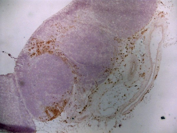
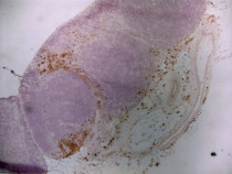
ARG23390 anti-CD163 antibody [ED2] IHC-Fr image
Immunohistochemistry: Acetone fixed, cryostat sectioned Rat spleen stained with ARG23390 anti-CD163 antibody [ED2].
-
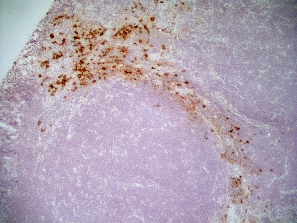
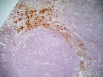
ARG23390 anti-CD163 antibody [ED2] IHC-Fr image
Immunohistochemistry: Rat lymph node cryosection stained with ARG23390 anti-CD163 antibody [ED2] followed by HRP-conjugated Goat anti Mouse IgG1 as a detection reagent. (Low power).
-
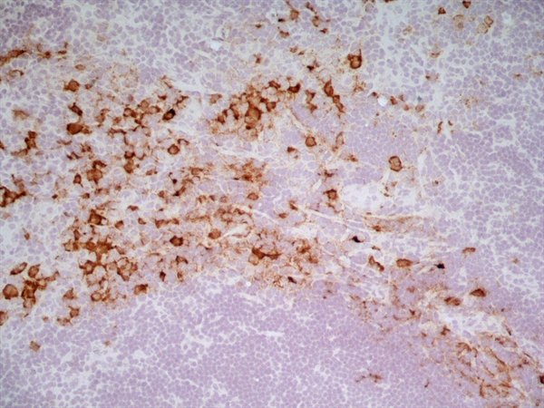
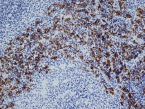
ARG23390 anti-CD163 antibody [ED2] IHC-Fr image
Immunohistochemistry: Rat lymph node cryosection stained with ARG23390 anti-CD163 antibody [ED2] followed by HRP-conjugated Goat anti Mouse IgG1 as a detection reagent. (Medium power).
-
ARG23390 anti-CD163 antibody [ED2] IHC-Fr image
Immunohistochemistry: Rat lymph node cryosection stained with ARG23390 anti-CD163 antibody [ED2] followed by HRP-conjugated Goat anti Mouse IgG1 as a detection reagent. (Medium power).
-
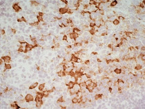
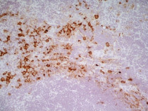
ARG23390 anti-CD163 antibody [ED2] IHC-Fr image
Immunohistochemistry: Rat lymph node cryosection stained with ARG23390 anti-CD163 antibody [ED2] followed by HRP-conjugated Goat anti Mouse IgG1 as a detection reagent. (High power).
-
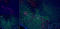
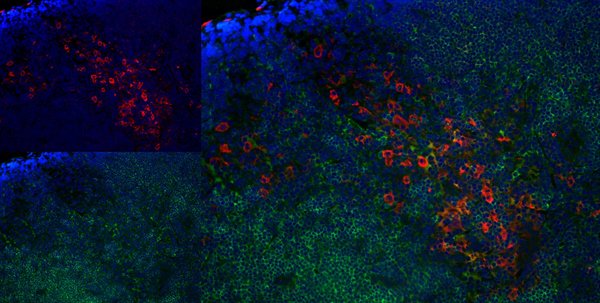
ARG23390 anti-CD163 antibody [ED2] IHC-Fr image
Immunohistochemistry: Rat lymph node cryosection stained with ARG23390 anti-CD163 antibody [ED2], red in A and Mouse anti Rat CD8, green in B. C is the merged image with nuclei counter-stained blue using DAPI. (Low power).
-

ARG23390 anti-CD163 antibody [ED2] IHC-Fr image
Immunohistochemistry: Rat lymph node cryosection stained with ARG23390 anti-CD163 antibody [ED2], red in A and Mouse anti Rat CD8, green in B. C is the merged image with nuclei counter-stained blue using DAPI. (High power).