ARG40980
anti-CBX3 / HP1 gamma antibody
anti-CBX3 / HP1 gamma antibody for Flow cytometry,ICC/IF,IHC-Frozen sections,IHC-Formalin-fixed paraffin-embedded sections,Western blot and Human,Mouse,Rat
Overview
| Product Description | Rabbit Polyclonal antibody recognizes CBX3 / HP1 gamma |
|---|---|
| Tested Reactivity | Hu, Ms, Rat |
| Tested Application | FACS, ICC/IF, IHC-Fr, IHC-P, WB |
| Host | Rabbit |
| Clonality | Polyclonal |
| Isotype | IgG |
| Target Name | CBX3 / HP1 gamma |
| Antigen Species | Human |
| Immunogen | Recombinant protein corresponding to A2-Q183 of Human CBX3 / HP1 gamma. |
| Conjugation | Un-conjugated |
| Alternate Names | HP1-GAMMA; Chromobox protein homolog 3; HP1Hs-gamma; HP1 gamma; HECH; Heterochromatin protein 1 homolog gamma; Modifier 2 protein |
Application Instructions
| Application Suggestion |
|
||||||||||||
|---|---|---|---|---|---|---|---|---|---|---|---|---|---|
| Application Note | IHC-P: Antigen Retrieval: Heat mediation was performed in Citrate buffer (pH 6.0) for 20 min. * The dilutions indicate recommended starting dilutions and the optimal dilutions or concentrations should be determined by the scientist. |
Properties
| Form | Liquid |
|---|---|
| Purification | Affinity purification with immunogen. |
| Buffer | 0.2% Na2HPO4, 0.9% NaCl, 0.05% Sodium azide and 4% Trehalose. |
| Preservative | 0.05% Sodium azide |
| Stabilizer | 4% Trehalose |
| Concentration | 0.5 mg/ml |
| Storage Instruction | For continuous use, store undiluted antibody at 2-8°C for up to a week. For long-term storage, aliquot and store at -20°C or below. Storage in frost free freezers is not recommended. Avoid repeated freeze/thaw cycles. Suggest spin the vial prior to opening. The antibody solution should be gently mixed before use. |
| Note | For laboratory research only, not for drug, diagnostic or other use. |
Bioinformation
| Database Links | |
|---|---|
| Gene Symbol | CBX3 |
| Gene Full Name | chromobox homolog 3 |
| Background | At the nuclear envelope, the nuclear lamina and heterochromatin are adjacent to the inner nuclear membrane. The protein encoded by this gene binds DNA and is a component of heterochromatin. This protein also can bind lamin B receptor, an integral membrane protein found in the inner nuclear membrane. The dual binding functions of the encoded protein may explain the association of heterochromatin with the inner nuclear membrane. This protein binds histone H3 tails methylated at Lys-9 sites. This protein is also recruited to sites of ultraviolet-induced DNA damage and double-strand breaks. Two transcript variants encoding the same protein but differing in the 5' UTR, have been found for this gene.[provided by RefSeq, Mar 2011] |
| Function | Seems to be involved in transcriptional silencing in heterochromatin-like complexes. Recognizes and binds histone H3 tails methylated at 'Lys-9', leading to epigenetic repression. May contribute to the association of the heterochromatin with the inner nuclear membrane through its interaction with lamin B receptor (LBR). Involved in the formation of functional kinetochore through interaction with MIS12 complex proteins. Contributes to the conversion of local chromatin to a heterochromatin-like repressive state through H3 'Lys-9' trimethylation, mediates the recruitment of the mehtyltransferases SUV39H1 and/or SUV39H2 by the PER complex to the E-box elements of the circadian target genes such as PER2 itself or PER1. [UniProt] |
| Cellular Localization | Nucleus. Note=Associates with euchromatin and is largely excluded from constitutive heterochromatin. May be associated with microtubules and mitotic poles during mitosis (Potential). [UniProt] |
| Highlight | Related products: CBX3 antibodies; CBX3 Duos / Panels; Anti-Rabbit IgG secondary antibodies; Related news: Senescence Marker Antibody Panel is launched |
| Calculated MW | 21 kDa |
| PTM | Phosphorylated by PIM1. Phosphorylated during interphase and possibly hyper-phosphorylated during mitosis. [UniProt] |
Images (11) Click the Picture to Zoom In
-
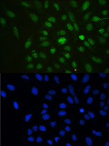
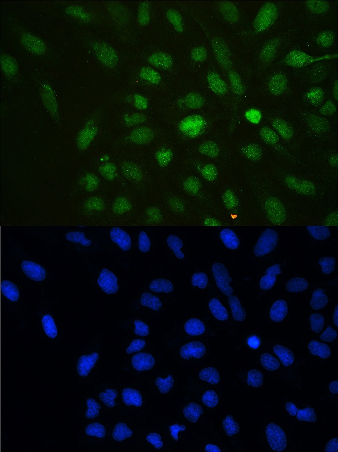
ARG40980 anti-CBX3 / HP1 gamma antibody ICC/IF image
Immunofluorescence: U2OS cells stained with ARG40980 anti-CBX3 / HP1 gamma antibody (green) at 2 µg/ml, overnight at 4°C. DAPI (blue) for nuclear staining.
-
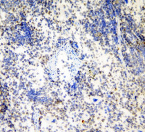
ARG40980 anti-CBX3 / HP1 gamma antibody IHC-Fr image
Immunohistochemistry: Frozen section of Human placenta tissue. The tissue section was blocked with 10% goat serum. The tissue section was then stained with ARG40980 anti-CBX3 / HP1 gamma antibody at 1 µg/ml dilution, overnight at 4°C.
-
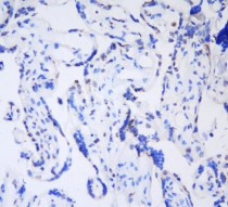
ARG40980 anti-CBX3 / HP1 gamma antibody IHC-P image
Immunohistochemistry: Paraffin-embedded Human mammary cancer tissue. Antigen Retrieval: Heat mediation was performed in Citrate buffer (pH 6.0, epitope retrieval solution) for 20 min. The tissue section was blocked with 10% goat serum. The tissue section was then stained with ARG40980 anti-CBX3 / HP1 gamma antibody at 1 µg/ml dilution, overnight at 4°C.
-
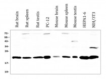
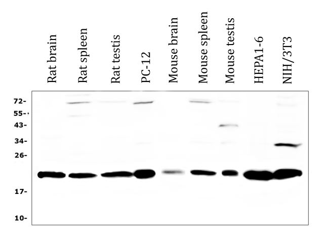
ARG40980 anti-CBX3 / HP1 gamma antibody WB image
Western blot: 50 µg of samples under reducing conditions. Rat brain, Rat spleen, Rat testis, PC-12, Mouse brain, Mouse spleen, Mouse testis, HEPA1-6 and NIH/3T3 cell lysates stained with ARG40980 anti-CBX3 / HP1 gamma antibody at 0.5 µg/ml, overnight at 4°C.
-
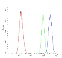
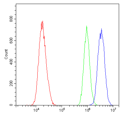
ARG40980 anti-CBX3 / HP1 gamma antibody FACS image
Flow Cytometry: A431 cells were blocked with 10% normal goat serum and then stained with ARG40980 anti-CBX3 / HP1 gamma antibody (blue) at 1 µg/10^6 cells for 30 min at 20°C, followed by incubation with DyLight®488 labelled secondary antibody. Isotype control antibody (green) was Rabbit IgG (1 µg/10^6 cells) used under the same conditions. Unlabelled sample (red) was also used as a control.
-
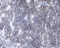
ARG40980 anti-CBX3 / HP1 gamma antibody IHC-Fr image
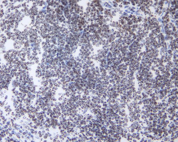
Immunohistochemistry: Frozen section of Rat spleen tissue. The tissue section was blocked with 10% goat serum. The tissue section was then stained with ARG40980 anti-CBX3 / HP1 gamma antibody at 1 µg/ml dilution, overnight at 4°C.
-
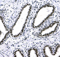
ARG40980 anti-CBX3 / HP1 gamma antibody IHC-P image
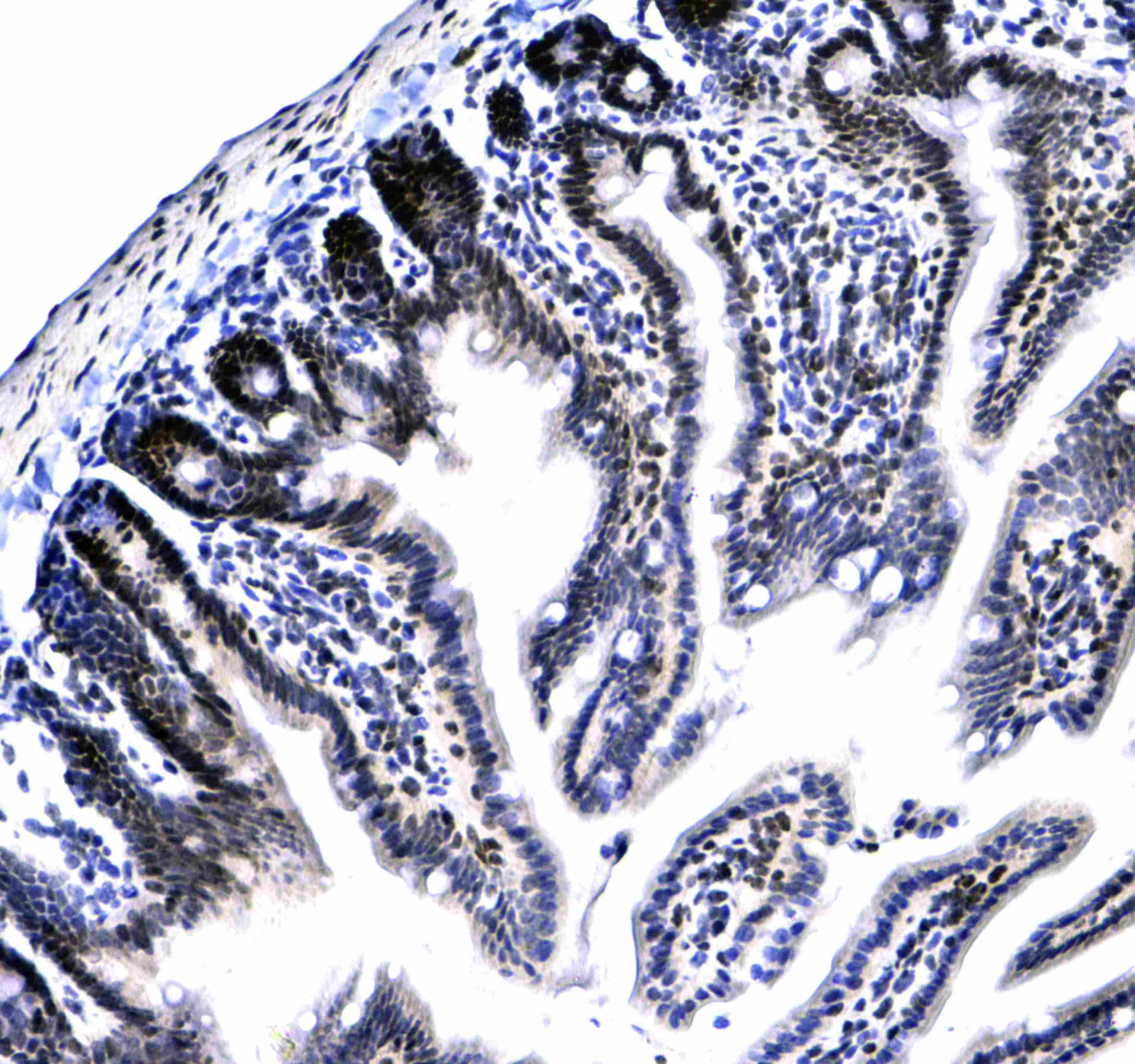
Immunohistochemistry: Paraffin-embedded Human rectal cancer tissue. Antigen Retrieval: Heat mediation was performed in Citrate buffer (pH 6.0, epitope retrieval solution) for 20 min. The tissue section was blocked with 10% goat serum. The tissue section was then stained with ARG40980 anti-CBX3 / HP1 gamma antibody at 1 µg/ml dilution, overnight at 4°C.
-
ARG40980 anti-CBX3 / HP1 gamma antibody IHC-Fr image
Immunohistochemistry: Frozen section of Mouse spleen tissue. The tissue section was blocked with 10% goat serum. The tissue section was then stained with ARG40980 anti-CBX3 / HP1 gamma antibody at 1 µg/ml dilution, overnight at 4°C.
-
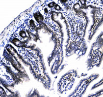
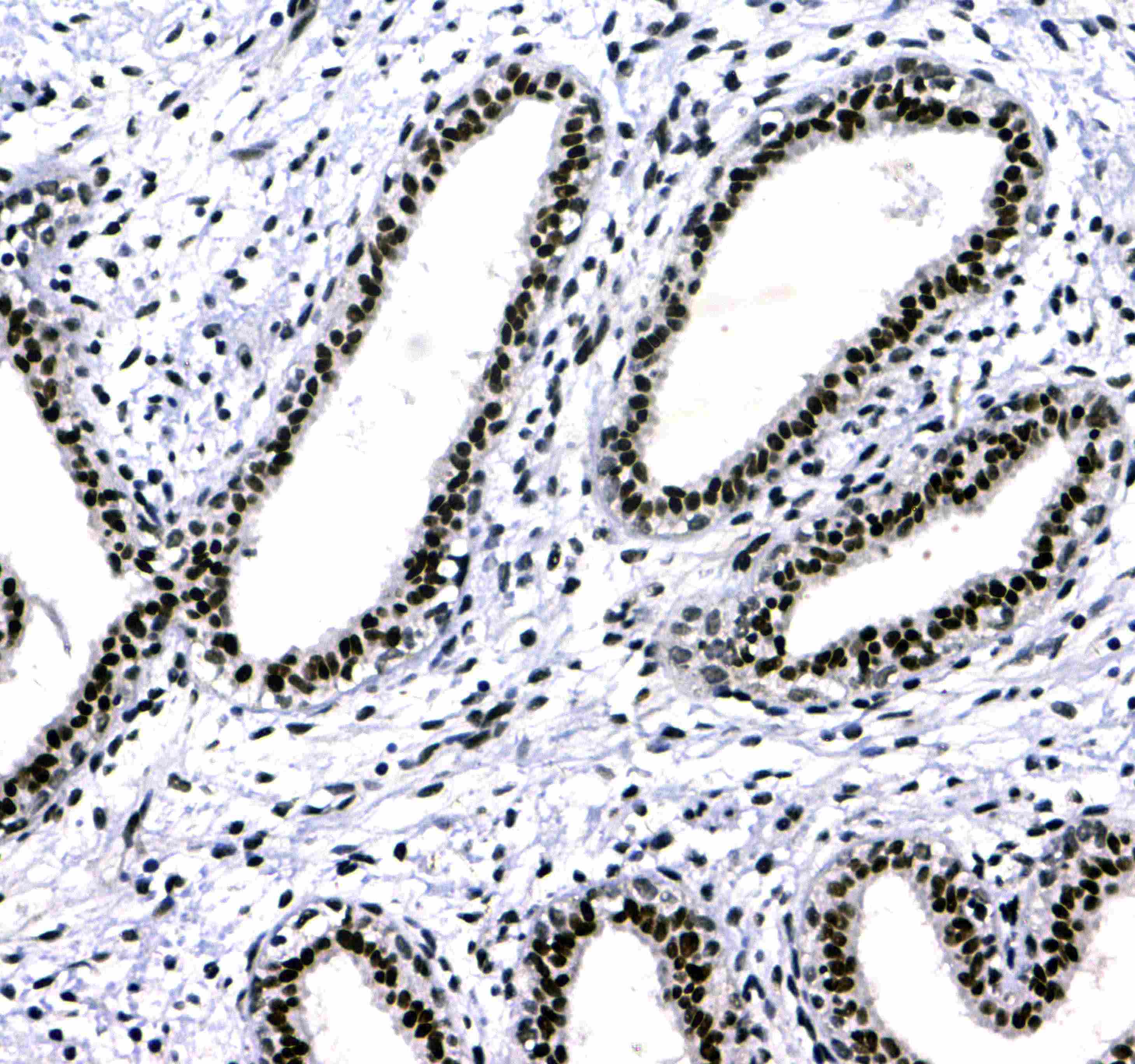
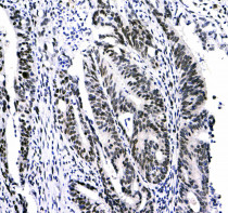
ARG40980 anti-CBX3 / HP1 gamma antibody IHC-P image
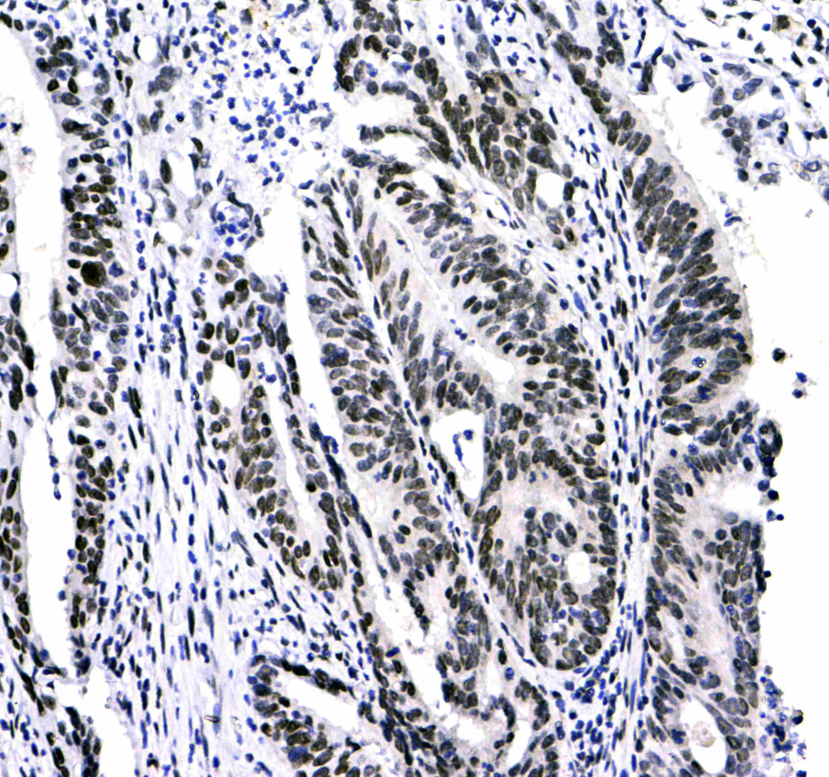
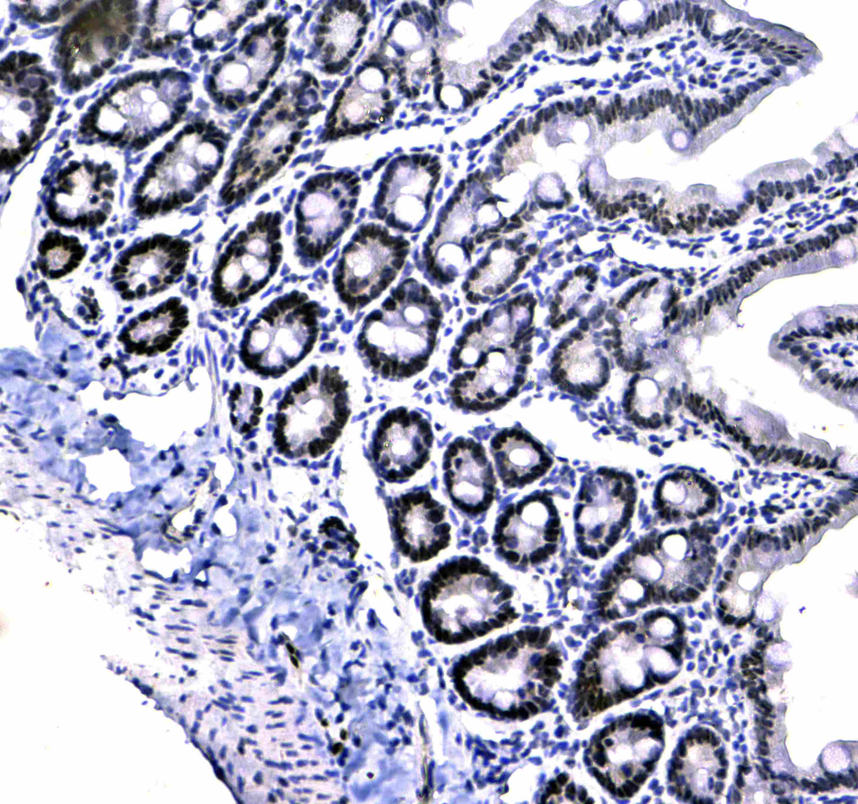
Immunohistochemistry: Paraffin-embedded Mouse small intestine tissue. Antigen Retrieval: Heat mediation was performed in Citrate buffer (pH 6.0, epitope retrieval solution) for 20 min. The tissue section was blocked with 10% goat serum. The tissue section was then stained with ARG40980 anti-CBX3 / HP1 gamma antibody at 1 µg/ml dilution, overnight at 4°C.
-
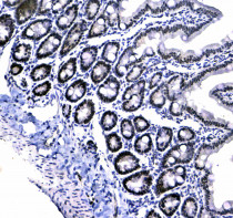
ARG40980 anti-CBX3 / HP1 gamma antibody IHC-P image
Immunohistochemistry: Paraffin-embedded Rat small intestine tissue. Antigen Retrieval: Heat mediation was performed in Citrate buffer (pH 6.0, epitope retrieval solution) for 20 min. The tissue section was blocked with 10% goat serum. The tissue section was then stained with ARG40980 anti-CBX3 / HP1 gamma antibody at 1 µg/ml dilution, overnight at 4°C.
-
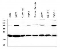
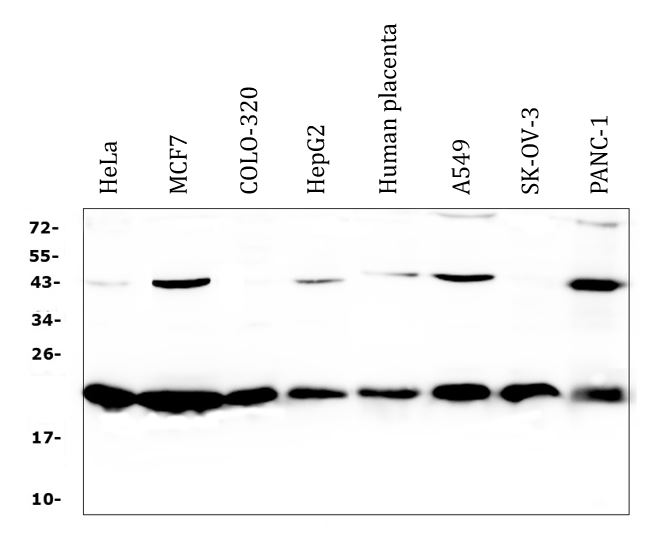
ARG40980 anti-CBX3 / HP1 gamma antibody WB image
Western blot: 50 µg of samples under reducing conditions. HeLa, MCF7, COLO-320, HepG2, Human placenta, A549, SK-OV-3 and PANC-1 cell lysates stained with ARG40980 anti-CBX3 / HP1 gamma antibody at 0.5 µg/ml, overnight at 4°C.