ARG42102
anti-BMP1 antibody
anti-BMP1 antibody for IHC-Formalin-fixed paraffin-embedded sections,Western blot and Human,Mouse,Rat
Overview
| Product Description | Goat Polyclonal antibody recognizes BMP1 |
|---|---|
| Tested Reactivity | Hu, Ms, Rat |
| Predict Reactivity | Cow, Dog, Pig |
| Tested Application | IHC-P, WB |
| Specificity | This antibody is expected to recognize both reported isoforms (NP_001190.1; NP_006120.1). |
| Host | Goat |
| Clonality | Polyclonal |
| Isotype | IgG |
| Target Name | BMP1 |
| Antigen Species | Human |
| Immunogen | Synthetic peptide around the internal region of Human BMP1. (C-DTIVPKYEVNGVK) (NP_001190.1; NP_006120.1) |
| Conjugation | Un-conjugated |
| Alternate Names | OI13; Procollagen C-proteinase; PCP; PCOLC; PCP2; BMP-1; Bone morphogenetic protein 1; Mammalian tolloid protein; TLD; EC 3.4.24.19; mTld |
Application Instructions
| Application Suggestion |
|
||||||
|---|---|---|---|---|---|---|---|
| Application Note | WB: Recommend incubate at RT for 1h. IHC-P: Antigen Retrieval: Steam tissue section in Citrate buffer (pH 6.0). * The dilutions indicate recommended starting dilutions and the optimal dilutions or concentrations should be determined by the scientist. |
||||||
| Observed Size | ~ 110 kDa (Human) |
Properties
| Form | Liquid |
|---|---|
| Purification | Affinity purified |
| Buffer | Tris saline (pH 7.3), 0.02% Sodium azide and 0.5% BSA. |
| Preservative | 0.02% Sodium azide |
| Stabilizer | 0.5% BSA |
| Concentration | 0.5 mg/ml |
| Storage Instruction | For continuous use, store undiluted antibody at 2-8°C for up to a week. For long-term storage, aliquot and store at -20°C or below. Storage in frost free freezers is not recommended. Avoid repeated freeze/thaw cycles. Suggest spin the vial prior to opening. The antibody solution should be gently mixed before use. |
| Note | For laboratory research only, not for drug, diagnostic or other use. |
Bioinformation
| Database Links | |
|---|---|
| Gene Symbol | BMP1 |
| Gene Full Name | bone morphogenetic protein 1 |
| Background | This gene encodes a protein that is capable of inducing formation of cartilage in vivo. Although other bone morphogenetic proteins are members of the TGF-beta superfamily, this gene encodes a protein that is not closely related to other known growth factors. This gene is expressed as alternatively spliced variants that share an N-terminal protease domain but differ in their C-terminal region. [provided by RefSeq, Aug 2008] |
| Function | Cleaves the C-terminal propeptides of procollagen I, II and III. Induces cartilage and bone formation. May participate in dorsoventral patterning during early development by cleaving chordin (CHRD). Responsible for the proteolytic activation of lysyl oxidase LOX. [UniProt] |
| Cellular Localization | Golgi apparatus, trans-Golgi network. Secreted, extracellular space, extracellular matrix. Note=Co-localizes with POSTN in the Golgi. [UniProt] |
| Calculated MW | 111 kDa (Human) |
| PTM | Proteolytically activated in the trans-Golgi network by furin-like/paired basic proprotein convertases, cleavage is not required for secretion. [UniProt] |
Images (5) Click the Picture to Zoom In
-
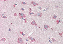
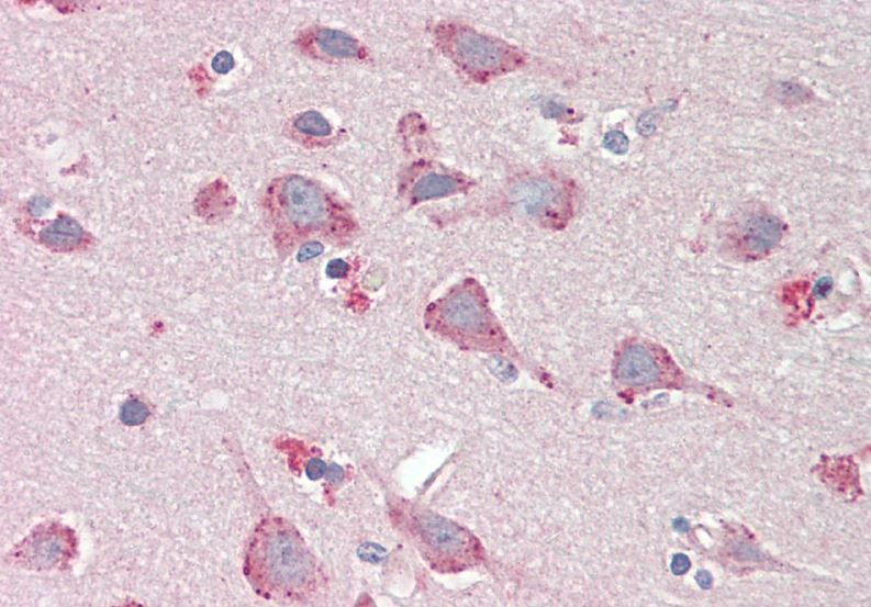
ARG42102 anti-BMP1 antibody IHC-P image
Immunohistochemistry: Paraffin-embedded Human cortex tissue. Antigen Retrieval: Steam tissue section in Citrate buffer (pH 6.0). The tissue section was stained with ARG42102 anti-BMP1 antibody at 5 µg/ml dilution.
-

ARG42102 anti-BMP1 antibody WB image
Western blot: 35 µg of Human heart and Human kidney lysates (in RIPA buffer) stained with ARG42102 anti-BMP1 antibody at 1 µg/ml dilution and incubated at RT for 1 hour.
-

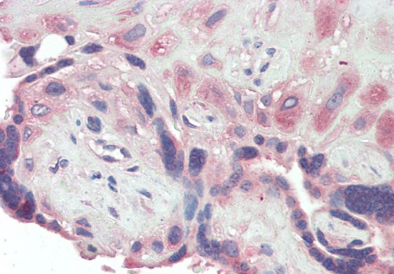
ARG42102 anti-BMP1 antibody IHC-P image
Immunohistochemistry: Paraffin-embedded Human placenta tissue. Antigen Retrieval: Steam tissue section in Citrate buffer (pH 6.0). The tissue section was stained with ARG42102 anti-BMP1 antibody at 5 µg/ml dilution.
-
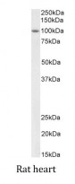
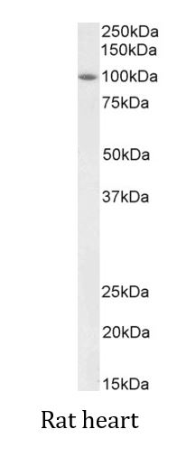
ARG42102 anti-BMP1 antibody WB image
Western blot: 35 µg of Rat heart lysate (in RIPA buffer) stained with ARG42102 anti-BMP1 antibody at 2 µg/ml dilution and incubated at RT for 1 hour.
-
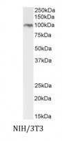
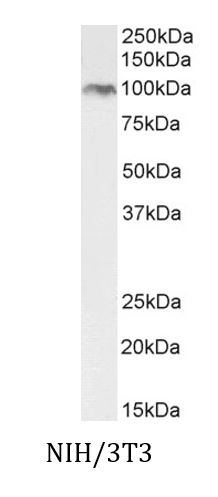
ARG42102 anti-BMP1 antibody WB image
Western blot: 35 µg of NIH/3T3 cell lysate (in RIPA buffer) stained with ARG42102 anti-BMP1 antibody at 2 µg/ml dilution and incubated at RT for 1 hour.