ARG58201
anti-BDNF antibody
anti-BDNF antibody for Flow cytometry,IHC-Formalin-fixed paraffin-embedded sections,Western blot and Human,Mouse,Rat
Overview
| Product Description | Rabbit Polyclonal antibody recognizes BDNF |
|---|---|
| Tested Reactivity | Hu, Ms, Rat |
| Tested Application | FACS, IHC-P, WB |
| Host | Rabbit |
| Clonality | Polyclonal |
| Isotype | IgG |
| Target Name | BDNF |
| Antigen Species | Human |
| Immunogen | Partial recombinant protein corresponding to aa. 129-247 of Human BDNF. |
| Conjugation | Un-conjugated |
| Alternate Names | Brain-derived neurotrophic factor; Abrineurin; BULN2; ANON2; BDNF |
Application Instructions
| Application Suggestion |
|
||||||||
|---|---|---|---|---|---|---|---|---|---|
| Application Note | IHC-P: Antigen Retrieval: Steam tissue section in Citrate buffer (pH 6.0) for 20 min followed by cooling at RT. * The dilutions indicate recommended starting dilutions and the optimal dilutions or concentrations should be determined by the scientist. |
||||||||
| Observed Size | ~ 28 kDa |
Properties
| Form | Liquid |
|---|---|
| Purification | Affinity purification with immunogen. |
| Buffer | PBS, 0.025% Sodium azide and 2.5% BSA. |
| Preservative | 0.025% Sodium azide |
| Stabilizer | 2.5% BSA |
| Concentration | 0.5 mg/ml |
| Storage Instruction | For continuous use, store undiluted antibody at 2-8°C for up to a week. For long-term storage, aliquot and store at -20°C or below. Storage in frost free freezers is not recommended. Avoid repeated freeze/thaw cycles. Suggest spin the vial prior to opening. The antibody solution should be gently mixed before use. |
| Note | For laboratory research only, not for drug, diagnostic or other use. |
Bioinformation
| Database Links | |
|---|---|
| Gene Symbol | BDNF |
| Gene Full Name | brain-derived neurotrophic factor |
| Background | The protein encoded by this gene is a member of the nerve growth factor family. It is induced by cortical neurons, and is necessary for survival of striatal neurons in the brain. Expression of this gene is reduced in both Alzheimer's and Huntington disease patients. This gene may play a role in the regulation of stress response and in the biology of mood disorders. Multiple transcript variants encoding distinct isoforms have been described for this gene. [provided by RefSeq, Jan 2009] |
| Function | During development, promotes the survival and differentiation of selected neuronal populations of the peripheral and central nervous systems. Participates in axonal growth, pathfinding and in the modulation of dendritic growth and morphology. Major regulator of synaptic transmission and plasticity at adult synapses in many regions of the CNS. The versatility of BDNF is emphasized by its contribution to a range of adaptive neuronal responses including long-term potentiation (LTP), long-term depression (LTD), certain forms of short-term synaptic plasticity, as well as homeostatic regulation of intrinsic neuronal excitability. [UniProt] |
| Cellular Localization | Cytoplasmic, secreted. [UniProt] |
| Highlight | Related products: BDNF antibodies; BDNF ELISA Kits; BDNF recombinant proteins; Anti-Rabbit IgG secondary antibodies; Related news: The role of HDGF in tumor angiogenesis |
| Calculated MW | 28 kDa |
| PTM | The propeptide is N-glycosylated and glycosulfated. Converted into mature BDNF by plasmin (PLG). [UniProt] |
Images (4) Click the Picture to Zoom In
-
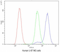
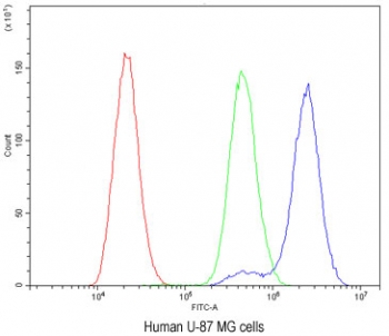
ARG58201 anti-BDNF antibody FACS image
Flow Cytometry: U-87 MG cells stained with ARG58201 anti-BDNF antibody at 1 µg/10^6 cells (blocked with Goat sera). Red: Cells alone, Green: Isotype control, Blue: Primary antibody.
-
ARG58201 anti-BDNF antibody IHC-P image
Immunohistochemistry: Paraffin-embedded Mouse brain tissue stained with ARG58201 anti-BDNF antibody. Antigen Retrieval: Steam tissue section in Citrate buffer (pH 6.0) for 20 min followed by cooling at RT.
-

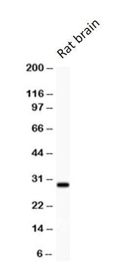
ARG58201 anti-BDNF antibody WB image
Western blot: Rat brain lysate stained with ARG58201 anti-BDNF antibody at 0.5 µg/ml dilution.
-
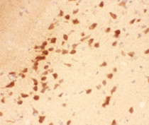
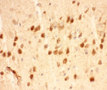
ARG58201 anti-BDNF antibody IHC-P image
Immunohistochemistry: Paraffin-embedded Rat brain tissue stained with ARG58201 anti-BDNF antibody. Antigen Retrieval: Steam tissue section in Citrate buffer (pH 6.0) for 20 min followed by cooling at RT.