ARG42648
anti-Arylsulfatase A antibody
anti-Arylsulfatase A antibody for Flow cytometry,ICC/IF,IHC-Formalin-fixed paraffin-embedded sections,Western blot and Human,Mouse,Rat
Overview
| Product Description | Rabbit Polyclonal antibody recognizes Arylsulfatase A |
|---|---|
| Tested Reactivity | Hu, Ms, Rat |
| Tested Application | FACS, ICC/IF, IHC-P, WB |
| Host | Rabbit |
| Clonality | Polyclonal |
| Isotype | IgG |
| Target Name | Arylsulfatase A |
| Antigen Species | Human |
| Immunogen | Synthetic peptide corresponding to aa. 454-482 of Human Arylsulfatase A. (QALKQLQLLKAQLDAAVTFGPSQVARGED) |
| Conjugation | Un-conjugated |
| Alternate Names | ASA; Cerebroside-sulfatase; EC 3.1.6.8; Arylsulfatase A; MLD |
Application Instructions
| Application Suggestion |
|
||||||||||
|---|---|---|---|---|---|---|---|---|---|---|---|
| Application Note | IHC-P: Antigen Retrieval: Heat mediation was performed in Citrate buffer (pH 6.0) for 20 min. * The dilutions indicate recommended starting dilutions and the optimal dilutions or concentrations should be determined by the scientist. |
||||||||||
| Observed Size | ~ 58 kDa |
Properties
| Form | Liquid |
|---|---|
| Purification | Affinity purification with immunogen. |
| Buffer | 0.2% Na2HPO4, 0.9% NaCl, 0.05% Sodium azide and 5% BSA. |
| Preservative | 0.05% Sodium azide |
| Stabilizer | 5% BSA |
| Concentration | 0.5 mg/ml |
| Storage Instruction | For continuous use, store undiluted antibody at 2-8°C for up to a week. For long-term storage, aliquot and store at -20°C or below. Storage in frost free freezers is not recommended. Avoid repeated freeze/thaw cycles. Suggest spin the vial prior to opening. The antibody solution should be gently mixed before use. |
| Note | For laboratory research only, not for drug, diagnostic or other use. |
Bioinformation
| Database Links | |
|---|---|
| Gene Symbol | ARSA |
| Gene Full Name | arylsulfatase A |
| Background | The protein encoded by this gene hydrolyzes cerebroside sulfate to cerebroside and sulfate. Defects in this gene lead to metachromatic leucodystrophy (MLD), a progressive demyelination disease which results in a variety of neurological symptoms and ultimately death. Alternatively spliced transcript variants have been described for this gene. [provided by RefSeq, Dec 2010] |
| Function | Hydrolyzes cerebroside sulfate. [UniProt] |
| Cellular Localization | Lysosome. [UniProt] |
| Calculated MW | 54 kDa |
| PTM | The conversion to 3-oxoalanine (also known as C-formylglycine, FGly), of a serine or cysteine residue in prokaryotes and of a cysteine residue in eukaryotes, is critical for catalytic activity. This post-translational modification is severely defective in multiple sulfatase deficiency (MSD). [UniProt] |
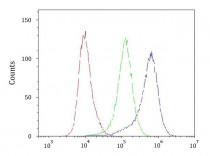
Images (6) Click the Picture to Zoom In
-

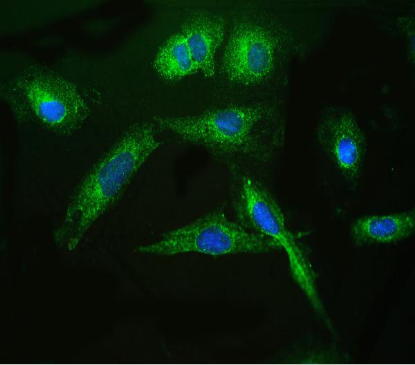
ARG42648 anti-Arylsulfatase A antibody ICC/IF image
Immunofluorescence: U2OS cells stained with ARG42648 anti-Arylsulfatase A antibody (green) at 2 µg/ml dilution, overnight at 4°C. DAPI (blue) for nuclear staining.
-

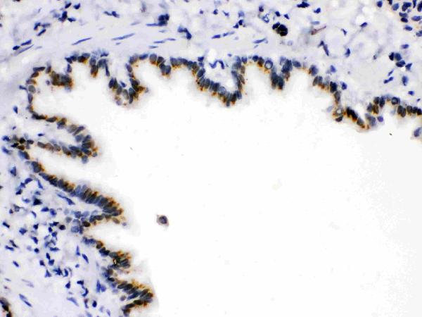
ARG42648 anti-Arylsulfatase A antibody IHC-P image
Immunohistochemistry: Paraffin-embedded Human mammary cancer tissue. Antigen Retrieval: Heat mediation was performed in Citrate buffer (pH 6.0) for 20 min. The tissue section was blocked with 10% goat serum. The tissue section was then stained with ARG42648 anti-Arylsulfatase A antibody at 1 µg/ml dilution, overnight at 4°C.
-

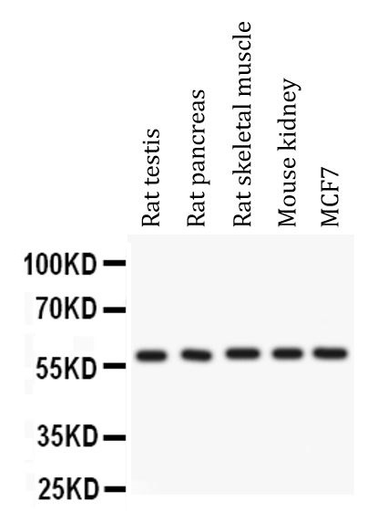
ARG42648 anti-Arylsulfatase A antibody WB image
Western blot: 50 µg of samples under reducing conditions. Rat testis, Rat pancreas, Rat skeletal muscle, Mouse kidney and MCF7 whole cell lysates stained with ARG42648 anti-Arylsulfatase A antibody at 0.5 µg/ml dilution, overnight at 4°C.
-
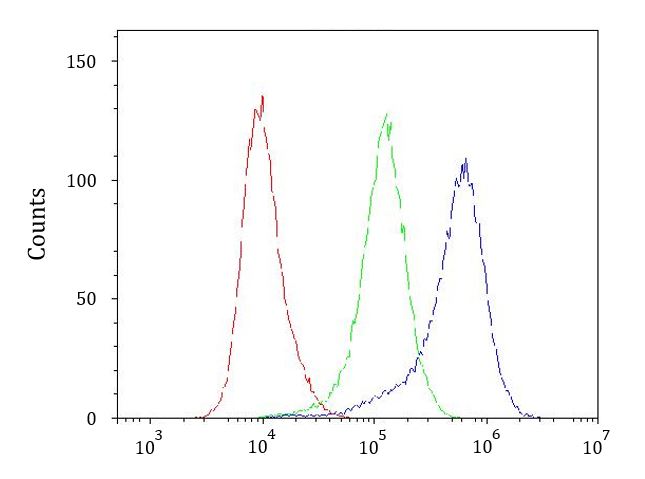
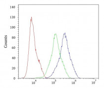
ARG42648 anti-Arylsulfatase A antibody FACS image
Flow Cytometry: HeLa cells were blocked with 10% normal goat serum and then stained with ARG42648 anti-Arylsulfatase A antibody (blue) at 1 µg/10^6 cells for 30 min at 20°C, followed by incubation with DyLight®488 labelled secondary antibody. Isotype control antibody (green) was Rabbit IgG (1 µg/10^6 cells) used under the same conditions. Unlabelled sample (red) was also used as a control.
-
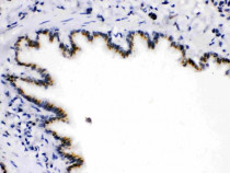
ARG42648 anti-Arylsulfatase A antibody IHC-P image
Immunohistochemistry: Paraffin-embedded Rat lung tissue. Antigen Retrieval: Heat mediation was performed in Citrate buffer (pH 6.0) for 20 min. The tissue section was blocked with 10% goat serum. The tissue section was then stained with ARG42648 anti-Arylsulfatase A antibody at 1 µg/ml dilution, overnight at 4°C.
-
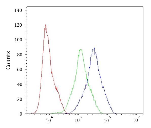
ARG42648 anti-Arylsulfatase A antibody FACS image
Flow Cytometry: PC-3 cells were blocked with 10% normal goat serum and then stained with ARG42648 anti-Arylsulfatase A antibody (blue) at 1 µg/10^6 cells for 30 min at 20°C, followed by incubation with DyLight®488 labelled secondary antibody. Isotype control antibody (green) was Rabbit IgG (1 µg/10^6 cells) used under the same conditions. Unlabelled sample (red) was also used as a control.