ARG22563
anti-ALIX antibody [3A9]
anti-ALIX antibody [3A9] for ELISA,ICC/IF,IHC-Formalin-fixed paraffin-embedded sections,Immunoprecipitation,Western blot and Human,Mouse,Rat,Xenopus
Overview
| Product Description | Mouse Monoclonal antibody [3A9] recognizes ALIX This antibody recognizes the apoptosis-linked gene-2 interacting protein X (ALIX), also known as Programmed cell death 6-interacting protein or Hp95. ALIX is a 868 amino acid ~90kDa cytoplasmic protein containing a single BRO1 domain implicated in endosomal targeting (Kim et al. 2005). ALIX was first reported to play a role in apoptosis dependent on its association with PDCD6 (Mahul-Mellier et al. 2006) but has subsequently shown to be involved in endocytosis (Matsuo et al. 2004), viral budding (Irie et al. 2008) and actin cytoskeletal assembly (Pan et al. 2006). Mouse anti Alix, clone 3A9 has been used extensively for the detection of ALIX by Western blotting and for the identification of an ALIX homologue in the sea urchin Paracentrotus lividus (Romancino et al. 2013). |
|---|---|
| Tested Reactivity | Hu, Ms, Rat, Xenopus |
| Tested Application | ELISA, ICC/IF, IHC-P, IP, WB |
| Host | Mouse |
| Clonality | Monoclonal |
| Clone | 3A9 |
| Isotype | IgG1 |
| Target Name | ALIX |
| Antigen Species | Human |
| Immunogen | Alix fusion protein. |
| Conjugation | Un-conjugated |
| Alternate Names | Programmed cell death 6-interacting protein; ALG-2-interacting protein 1; ALG-2-interacting protein X; DRIP4; PDCD6-interacting protein; Hp95; ALIX; HP95; AIP1 |
Application Instructions
| Application Suggestion |
|
||||||||||||
|---|---|---|---|---|---|---|---|---|---|---|---|---|---|
| Application Note | Immunohistology: This product does not require protein digestion pre-treatment of paraffin sections. * The dilutions indicate recommended starting dilutions and the optimal dilutions or concentrations should be determined by the scientist. |
Properties
| Form | Liquid |
|---|---|
| Purification | Purification with Protein G. |
| Buffer | PBS and 0.09% Sodium azide |
| Preservative | 0.09% Sodium azide |
| Concentration | 1 mg/ml |
| Storage Instruction | For continuous use, store undiluted antibody at 2-8°C for up to a week. For long-term storage, aliquot and store at -20°C or below. Storage in frost free freezers is not recommended. Avoid repeated freeze/thaw cycles. Suggest spin the vial prior to opening. The antibody solution should be gently mixed before use. |
| Note | For laboratory research only, not for drug, diagnostic or other use. |
Bioinformation
| Database Links | |
|---|---|
| Gene Symbol | PDCD6IP |
| Gene Full Name | programmed cell death 6 interacting protein |
| Background | This gene encodes a protein that functions within the ESCRT pathway in the abscission stage of cytokinesis, in intralumenal endosomal vesicle formation, and in enveloped virus budding. Studies using mouse cells have shown that overexpression of this protein can block apoptosis. In addition, the product of this gene binds to the product of the PDCD6 gene, a protein required for apoptosis, in a calcium-dependent manner. This gene product also binds to endophilins, proteins that regulate membrane shape during endocytosis. Overexpression of this gene product and endophilins results in cytoplasmic vacuolization, which may be partly responsible for the protection against cell death. Several alternatively spliced transcript variants encoding different isoforms have been found for this gene. Related pseudogenes have been identified on chromosome 15. [provided by RefSeq, Jan 2012] |
| Function | Class E VPS protein involved in concentration and sorting of cargo proteins of the multivesicular body (MVB) for incorporation into intralumenal vesicles (ILVs) that are generated by invagination and scission from the limiting membrane of the endosome. Binds to the phospholipid lysobisphosphatidic acid (LBPA) which is abundant in MVBs internal membranes. The MVB pathway appears to require the sequential function of ESCRT-O, -I,-II and -III complexes. The ESCRT machinery also functions in topologically equivalent membrane fission events, such as the terminal stages of cytokinesis and enveloped virus budding (HIV-1 and other lentiviruses). Appears to be an adapter for a subset of ESCRT-III proteins, such as CHMP4, to function at distinct membranes. Required for completion of cytokinesis. Involved in HIV-1 virus budding. Can replace TSG101 it its role of supporting HIV-1 release; this function implies the interaction with CHMP4B. May play a role in the regulation of both apoptosis and cell proliferation. [UniProt] |
| Highlight | Related products: Anti-Mouse IgG secondary antibodies; Related news: Detecting exosomal PD-L1 secreted by cancer cells |
| Calculated MW | 96 kDa |
| PTM | May be phosphorylated on tyrosine residues by activated PDGFRB. |
Images (1) Click the Picture to Zoom In
-
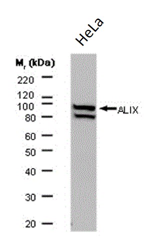
ARG22563 anti-ALIX antibody [3A9] WB image
Western blot: HeLa whole cell lysate stained with ARG22563 anti-ALIX antibody [3A9].