ARG30275
Pro-apoptotic Bcl2 protein Antibody Panel (BAX, BAK, Bid)
Component
| Cat No | Component Name | Host clonality | Reactivity | Application | Package |
|---|---|---|---|---|---|
| ARG54941 | anti-Bax antibody | Rabbit pAb | Hu, Ms | ICC/IF, IP, WB | 50 μg |
| ARG62367 | anti-Bak antibody | Rabbit pAb | Hu, Ms, Hm | IHC-P, WB | 100 μl |
| ARG65625 | anti-Bid antibody | Rabbit pAb | Hu | IHC-P, WB | 50 μl |
| ARG65351 | Goat anti-Rabbit IgG antibody (HRP) | Goat pAb | Rb | ELISA, IHC-P, WB | 50 μl |
Overview
| Product Description | A variety of physiological death signals trigger the genetically programmed pathway of apoptosis which is manifested in two major execution programs downstream of the death signal: the caspase pathway and organelle dysfunction. The BCL-2 family of proteins is known as an important gatekeeper to the apoptotic response. This group of structurally related proteins comprises pro-apoptotic and anti-apoptotic members that interact with one another. The pro-apoptotic factors, including BAX, BAK or BID promotes pore-forming effectors such that Cytochrome-C is released from the mitochondria and kick-start the caspase cleavage cascades. They, together with their opposing partner, BCL2, have become targets for cancer therapeutic interventions. Gross et al. 1999. Genes & Dev 13:1899-1911 |
|---|---|
| Target Name | Pro-apoptotic Bcl2 protein |
| Alternate Names | Pro-apoptotic Bcl2 protein antibody; Bax antibody; Bak antibody; Bid antibody |
Properties
| Storage Instruction | For continuous use, store undiluted antibody at 2-8°C for up to a week. For long-term storage, aliquot and store at -20°C or below. Storage in frost free freezers is not recommended. Avoid repeated freeze/thaw cycles. Suggest spin the vial prior to opening. The antibody solution should be gently mixed before use. |
|---|---|
| Note | For laboratory research only, not for drug, diagnostic or other use. |
Bioinformation
| Gene Full Name | Antibody Panel for Pro-apoptotic Bcl2 protein (BAX, BAK, Bid) |
|---|---|
| Highlight | Related Product: anti-Bax antibody; anti-Bak antibody; anti-Bid antibody; |
Images (10) Click the Picture to Zoom In
-
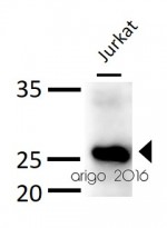
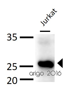
ARG62367 anti-Bak antibody WB image
Western blot: 30 µg of Jurkat cell lysate stained with ARG62367 anti-Bak antibody at 1:500 dilution.
-
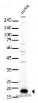
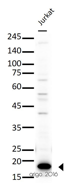
ARG65625 anti-Bid antibody WB image
Western blot: 30 µg of Jurkat cell lysate stained with ARG65625 anti-Bid antibody at 1:500 dilution.
-

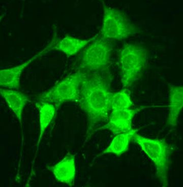
ARG54941 anti-Bax antibody ICC/IF image
Immunofluorescence: NIH/3T3 cells stained with ARG54941 anti-Bax antibody at 1:100 dilution.
-

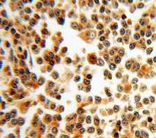
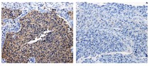
ARG54941 anti-Bax antibody IHC-P image
Immunohistochemistry: paraffin-embedded Lymphoma stained with ARG54941 anti-Bax antibody.
-
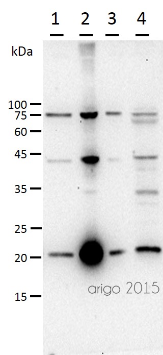
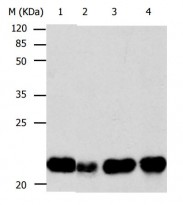
ARG54941 anti-Bax antibody WB image
Western blot: 30 µg of 1) HeLa, 2) HepG2, 3) 293T, and 4) Mouse ovary lysate stained with ARG54941 anti-Bax antibody at 1:500 dilution.
-
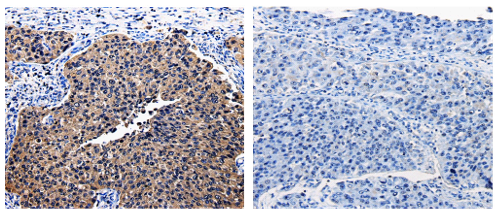
ARG65625 anti-Bid antibody IHC-P image
Immunohistochemistry: Paraffin-embedded Human renal cancer tissue stained with ARG65625 anti-Bid antibody (left) at 1:25 dilution, or the same antibody preincubated with antigen (right). (Original magnification: X200)
-
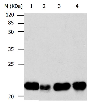
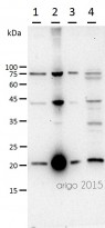
ARG65625 anti-Bid antibody WB image
Western Blot: 40 μg of lysates from: 1) Human fetal brain tissue, 2) Human liver tissue, 3) HT29 cells, 4) HepG2 cells stained with ARG65625 anti-Bid antibody at 1/260 dilution. Exposure time: 10 seconds.
-

ARG65351 Goat anti-Rabbit IgG antibody (HRP) WB image
Western blot: Rat placental stained with ARG57589 anti-MTNR1A antibody at 1:1000 dilution, ARG65351 Goat anti-Rabbit IgG antibody (HRP) at 1:5000 dilution.
From Jinzhi Li et al. J Reprod Immunol. (2023), doi: 10.1016/j.jri.2023.104166, Fig. 2.B.
-

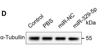
ARG65351 Goat anti-Rabbit IgG antibody (HRP) WB image
Western blot: Mouse retina stained with ARG65693 anti-alpha Tubulin antibody and ARG65351 Goat anti-Rabbit IgG antibody (HRP)
From Xiaoyuan Ye et al. Mol Ther Nucleic Acids. (2024), doi: 10.1016/j.omtn.2024.102209, Fig. 5.D.
-
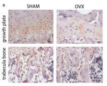
ARG65351 Goat anti-Rabbit IgG antibody (HRP) IHC-P image
From Yu-Qian Song et al. J Mol Med (Berl) (2022), doi: 10.1007/s00109-021-02165-0, Fig. 5.c.