ARG30273
LC3A and LC3B Antibody Duo
Component
| Cat No | Component Name | Host clonality | Reactivity | Application | Package |
|---|---|---|---|---|---|
| ARG51300 | anti-LC3A antibody | Rabbit pAb | Hu, Ms, Rat | ICC/IF, IHC-P, WB | 50 μl |
| ARG55799 | anti-LC3B antibody | Rabbit pAb | Hu, Ms, Rat | ICC/IF, IHC-P, WB | 50 μl |
Overview
| Product Description | Autophagy consists of a destructive mechanism that disassemble unnecessary or dysfunctional cellular components through a regulated process. Targeted cellular constituents are enclosed by double-membraned vesicle called autophagosome. LC3s (MAP1-LC3A, B and C) are structural proteins of autophagosomal membranes, widely used as biomarkers of autophagy. They are processed from their precursors and bound to PE to form autophagosome before being fused to lysosome and breakdown the enclosed components. In the cytoplasm, there was a minimal co-localization between LC3A and B staining, suggesting that the relevant autophagosomes are formed by only one out of the two LC3 proteins. Koukourakis M. et al. 2015. Plos One 17(10) Related news: Keap1-Nrf2-ARE antibody panel is launched Freeze! Scientists found a way to stop tumor migration Therapeutic strategies against PDAC |
|---|---|
| Target Name | LC3A and LC3B |
| Alternate Names | LC3A and LC3B antibody; Microtubule-associated protein 1 light chain 3 alpha and Microtubule-associated protein 1 light chain 3 beta antibody; LC3A antibody; LC3B antibody |
Properties
| Storage Instruction | For continuous use, store undiluted antibody at 2-8°C for up to a week. For long-term storage, aliquot and store at -20°C or below. Storage in frost free freezers is not recommended. Avoid repeated freeze/thaw cycles. Suggest spin the vial prior to opening. The antibody solution should be gently mixed before use. |
|---|---|
| Note | For laboratory research only, not for drug, diagnostic or other use. |
Bioinformation
| Gene Full Name | Microtubule-associated protein 1 light chain 3 alpha (LC3A) and Microtubule-associated protein 1 light chain 3 beta (LC3B) Antibody Duo |
|---|---|
| Highlight | Related products: LC3 antibodies; LC3 Duos / Panels; |
Images (9) Click the Picture to Zoom In
-
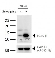
ARG51300 anti-LC3A antibody WB image
Western blot: 30 µg of HeLa untreated or treated with CQ (50 µM) and stained with ARG51300 anti-LC3A antibody at 1:1000 dilution.
-
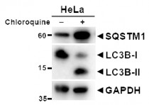
ARG55799 anti-LC3B antibody WB image (Customer's Feedback)
Western blot: HeLa cells untreated or treated with Chloroquine. 20 µg of cell lysates stained with ARG55040 anti-SQSTM1 / p62 antibody and ARG55799 anti-LC3B antibody at 1:1000 dilution.
An anti-GAPDH antibody used as a positive control.
-
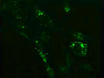
ARG55799 anti-LC3B antibody ICC/IF image
Immunofluorescence: NIH/3T3 cells treated with Chloroquine (50 μM, 37°C for 20 hours). Cells were stained with ARG55799 anti-LC3B antibody at 1:100 dilution.
-
ARG55799 anti-LC3B antibody IHC-P image
Immunohistochemistry: Paraffin-embedded Rat kidney stained with ARG55799 anti-LC3B antibody at 1:100 dilution.
-
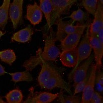
ARG51300 anti-LC3A antibody ICC/IF image
Immunofluorescence: methanol-fixed HeLa cells stained with anti-LC3A antibody ARG51300.
-
ARG51300 anti-LC3A antibody WB image
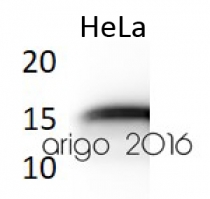
Western blot: 30 µg of HeLa cell lysate stained with ARG51300 anti-LC3A antibody at 1:500 dilution.
-
ARG51300 anti-LC3A antibody WB image
Western blot: 30 µg of HeLa cells untreated or treated with Chloroquine at 50 µM or 100 µM (18 hr). The blots stained with ARG51300 anti-LC3A antibody at 1:500 dilution.
-
ARG55799 anti-LC3B antibody WB image
Western blot: 30 µg of HeLa untreated or treated with Chloroquine at 50 µM, 100 µM (18 hr). The bolts stained with ARG55799 anti-LC3B antibody at 1:1000 dilution.
-
ARG55799 anti-LC3B antibody WB image
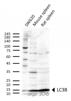
Western blot: 30 µg of SW620, Mouse spleen, and Rat spleen lysates stained with ARG55799 anti-LC3B antibody at 1:1000 dilution.
Specific References
