ARG30316
ER Marker Antibody Duo
Component
| Cat No | Component Name | Host clonality | Reactivity | Application | Package |
|---|---|---|---|---|---|
| ARG55123 | anti-Calreticulin antibody | Rabbit pAb | Hu, Ms, Rat | ICC/IF, IHC-P, WB | 50 μl |
| ARG20531 | anti-BiP / GRP78 antibody | Rabbit pAb | Hu, Ms, Rat, Bov, Dog, Fungi, Hm, Mk, Rb, Xenopus laevis | ICC/IF, IHC-FoFr , IHC-P, IP, WB | 50 μl |
Overview
| Product Description | Endoplasmic reticulum (ER) is a network of membrane-enclosed tubules and sacs (cisternae) that extends from the nuclear membrane throughout the cytoplasm. The ER lumen is the area enclosed by the ER membrane. ER is responsible for protein synthesis and folding, protein transport into the Golgi apparatus, and lipid metabolism. arigo's ER Marker Antibody Duo comprises an ER membrane marker Calreticulin antibody and an ER lumen marker BiP antibody. It is an excellent solution for labeling ER structures. |
|---|---|
| Target Name | ER Marker |
| Alternate Names | ER Marker antibody; BiP / GRP78 antibody; Calreticulin antibody |
Properties
| Storage Instruction | For continuous use, store undiluted antibody at 2-8°C for up to a week. For long-term storage, aliquot and store at -20°C or below. Storage in frost free freezers is not recommended. Avoid repeated freeze/thaw cycles. Suggest spin the vial prior to opening. The antibody solution should be gently mixed before use. |
|---|---|
| Note | For laboratory research only, not for drug, diagnostic or other use. |
Bioinformation
| Gene Full Name | Antibody Duo for ER Marker |
|---|---|
| Highlight | Related Product: anti-Calreticulin antibody; anti-BiP / GRP78 antibody; |
Images (10) Click the Picture to Zoom In
-
ARG55123 anti-Calreticulin antibody WB image
Western blot: 30 µg of HeLa, HT29, A549, MCF-7, HepG2, Jurkat, K562, Mouse ovary and Rat ovary lysates stained with ARG55123 anti-Calreticulin antibody at 1:500 dilution.
-
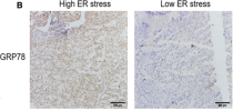
ARG20531 anti-BiP / GRP78 antibody IHC-P image
Immunohistochemistry: Human hepatocellular carcinoma stained with ARG20531 anti-BiP / GRP78 antibody at 1:500 dilution.
From Yue Zhu et al. Cancer Med. (2020), doi: 10.1002/cam4.2977, Fig. 5B.
-
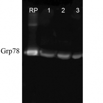
ARG20531 anti-BiP / GRP78 antibody WB image
Western blot: Human hepatocellular carcinoma stained with ARG20531 anti-BiP / GRP78 antibody at 1:500 dilution.
From Yue Zhu et al. Cancer Med. (2020), doi: 10.1002/cam4.2977, Fig. 5D.
-
ARG20531 anti-BiP / GRP78 antibody ICC/IF image
Immunofluorescence: Heat Shocked (42°C for 30 min) HeLa cells. Fixation: 2% Formaldehyde for 20 min at RT. Primary Antibody: ARG20531 anti-BiP / GRP78 antibody at 1:100 for 12 hours at 4°C. Secondary Antibody: FITC Goat anti-Rabbit (green) at 1:200 for 2 hours at RT. Counterstain: DAPI (blue) nuclear stain at 1:40000 for 2 hours at RT. Magnification: 100x. Left: DAPI (blue) nuclear stain. Middle: ARG20531 anti-BiP / GRP78 antibody. Right: Composite.
-
ARG20531 anti-BiP / GRP78 antibody IHC image
Immunohistochemistry: Formaldehdye-fixed and parrafin embedded sections of Mouse hippocampal tissue stained with ARG20531 anti-BiP / GRP78 antibody at 1:100 dilution. Tissue was counterstained using DAPI at a 1:1000 dilution in order to visualize the nuclei in the pyramidal cell layer. Images courtsey of Rachel Reith, NIH/NIMH.
-
ARG20531 anti-BiP / GRP78 antibody WB image
Western blot: Human, Dog, Mouse cell line lysates stained with ARG20531 anti-BiP / GRP78 antibody at 1:1000 dilution.
-
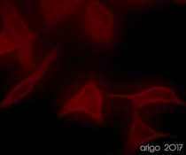
ARG55123 anti-Calreticulin antibody ICC/IF image
Immunofluorescence: 100% Methanol fixed (RT, 10 min) HeLa cells stained with ARG55123 anti-Calreticulin antibody (red) at 1:20 dilution.
Secondary antibody: ARG21917 Goat anti-Rabbit IgG antibody (TRITC)
-
ARG55123 anti-Calreticulin antibody ICC/IF image
Immunofluorescence: 100% Methanol fixed (RT, 10 min) HeLa cells stained with ARG55123 anti-Calreticulin antibody at 1:20 dilution. Left: primary antibody (red). Middle: DAPI (blue). Right: Merge.
Secondary antibody: ARG21917 Goat anti-Rabbit IgG antibody (TRITC)
-
ARG20531 anti-BiP / GRP78 antibody ICC/IF image
Immunofluorescence: Heat Shocked (42°C for 30 min) HeLa cells. Fixation: 2% Formaldehyde for 20 min at RT. Primary Antibody: ARG20531 anti-BiP / GRP78 antibody at 1:100 for 12 hours at 4°C. Secondary Antibody: APC Goat anti-Rabbit (red) at 1:200 for 2 hours at RT. Counterstain: DAPI (blue) nuclear stain at 1:40000 for 2 hours at RT. Magnification: 20x. Left: DAPI (blue) nuclear stain. Middle: ARG20531 anti-BiP / GRP78 antibody. Right: Composite.
-
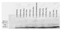
ARG20531 anti-BiP / GRP78 antibody WB image
Western blot: Rat brain, heart, kidney, liver, lung, pancreas, skeletal muscle, spleen, testes, thymus, H9C2 cell, Mouse NIH 3T3 and Pam212 cell lysates stained with ARG20531 anti-BiP / GRP78 antibody.
Specific References


