ARG30013
Colorectal Carcinoma Marker Antibody Duo (Colorectal carcinoma antibody, PKM2)
Cancer antibody; Controls and Markers antibody; Gene Regulation antibody; Metabolism antibody; Signaling Transduction antibody
Component
| Cat No | Component Name | Host clonality | Reactivity | Application | Package |
|---|---|---|---|---|---|
| ARG10083 | anti-Colorectal carcinoma antibody [Y94] | Mouse mAb | Hu | IHC-P | 50 μg |
| ARG61996 | anti-PKM1 / 2 antibody | Rabbit pAb | Hu, Ms, Rat | ICC/IF, IHC-P, WB | 50 μl |
Overview
| Product Description | Pyruvate kinase type M2 (PKM2) has been reported to be involved in aerobic glycolysis and cell growth in various tumors. In addition, in 2011 Tong J et al., reported that increasing PKM2 is also observed in sera and tissues from colorectal cancer patients with poor response to 5-FU. ARG30013 Colorectal Carcinoma Marker Duos, including antibody reacts PKM2 as a new Colorectal Carcinoma Marker and a Colorectal carcinoma antibody [Y94] which detects human colorectal carcinoma in tissue, but does not react with negative control normal adult tissues tested such as liver, colon, breast, stomach, lymph node, and kidney. This antibody Duo is useful for Colorectal Carcinoma study or PKM2 related cancer study. |
|---|---|
| Target Name | Colorectal Carcinoma Marker |
| Alternate Names | Colorectal Carcinoma Marker antibody; Colorectal carcinoma antibody; PKM1/2 antibody |
Properties
| Storage Instruction | For continuous use, store undiluted antibody at 2-8°C for up to a week. For long-term storage, aliquot and store at -20°C or below. Storage in frost free freezers is not recommended. Avoid repeated freeze/thaw cycles. Suggest spin the vial prior to opening. The antibody solution should be gently mixed before use. |
|---|---|
| Note | For laboratory research only, not for drug, diagnostic or other use. |
Bioinformation
| Gene Full Name | Antibody Duo for Colorectal Carcinoma Marker (Colorectal carcinoma antibody, PKM2) |
|---|---|
| Highlight | Related Product: anti-PKM1 / 2 antibody; |
| Research Area | Cancer antibody; Controls and Markers antibody; Gene Regulation antibody; Metabolism antibody; Signaling Transduction antibody |
Images (7) Click the Picture to Zoom In
-
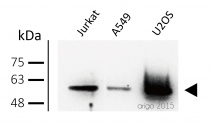
ARG61996 anti-PKM1/2 antibody WB image
Western blot: 30 µg of Jurkat, A549, and U2OS cell lysates stained with ARG61996 anti-PKM1/2 antibody at 1:500 dilution.
-
ARG61996 anti-PKM1/2 antibody ICC/IF image
Immunofluorescence: 100% Methanol fixed (RT, 10 min) HeLa cells stained with ARG61996 anti-PKM1/2 antibody at 1:20 dilution. Left: primary antibody (red). Middle: DAPI (blue). Right: Merge.
Secondary antibody: ARG21917 Goat anti-Rabbit IgG antibody (TRITC)
-
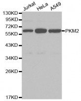
ARG61996 anti-PKM2 antibody WB image
Western Blot: extracts of various cell lines stained with anti-PKM2 antibody ARG61996.
-
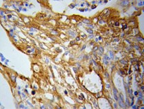
ARG61996 anti-PKM2 antibody IHC-P image
Immunohistochemistry: paraffin-embedded Lung cancer stained with anti-PKM2 antibody ARG61996.
-
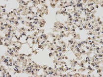
ARG61996 anti-PKM2 antibody IHC-P image
Immunohistochemistry: paraffin-embedded mouse lung stained with anti-PKM2 antibody ARG61996 at dilution of 1:100 (400x lens).
-
ARG61996 anti-PKM2 antibody IHC-P image
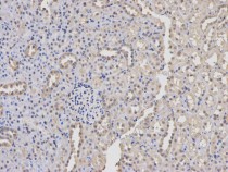
Immunohistochemistry: paraffin-embedded rat kidney stained with anti-PKM2 antibody ARG61996 at dilution of 1:100 (200x lens).
-
ARG61996 anti-PKM2 antibody ICC/IF image
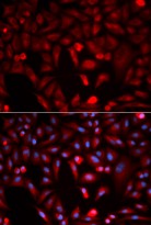
Immunofluorescence: HeLa cell stained with anti-PKM2 antibody ARG61996. Blue: DAPI for nuclear staining.
