ARG30314
Chondrogenesis Marker Antibody Panel
Component
| Cat No | Component Name | Host clonality | Reactivity | Application | Package |
|---|---|---|---|---|---|
| ARG57983 | anti-CDKN2A / p16INK4a antibody | Rabbit pAb | Hu | FACS, ICC/IF, IHC-P, IP, WB | 50 μl |
| ARG63341 | anti-FOXC1 antibody | Goat pAb | Hu | FACS, ICC/IF, IHC-P | 50 μg |
| ARG63332 | anti-FOXC2 antibody | Goat pAb | Hu | FACS, IHC-P, WB | 50 μg |
| ARG62852 | anti-CD44 antibody [MEM-263] | Mouse mAb | Hu, Dog, Pig | FACS, IHC-P, IP, WB | 50 μg |
| ARG65350 | Goat anti-Mouse IgG antibody (HRP) | Goat pAb | Ms | ELISA, IHC-P, WB | 50 μl |
| ARG65351 | Goat anti-Rabbit IgG antibody (HRP) | Goat pAb | Rb | ELISA, IHC-P, WB | 50 μl |
| ARG65352 | Donkey anti-Goat IgG antibody (HRP) | Donkey pAb | Goat | ELISA, IHC, WB | 50 μl |
Overview
| Product Description | Chondrogenesis is a process involving Mesenchymal Stem Cells (MSCc) condensation leading to chondroprogenitor cell differentiation. The main function of chondrogenensis is to generate cartilage during fetal development, and gradually replacing cartilage with bone at adulthood. Transcription factor SOX9, FOXC1 and FOXC2 are critical for driving chondrocyte differentiation. Hyaluronan receptor CD44 plays important role in cell-matrix interaction during chondrogenesis and matrix assembly. Hardinghan et al (2006). J Anat 209(4): 469-80 Wilm et al (2004). Dev Biol 271:176-189 Knudson CB (2003). Birth Defects Res C Embryo Today 69(2):174-96 |
|---|---|
| Target Name | Chondrogenesis Marker |
| Alternate Names | Chondrogenesis Marker antibody; CDKN2A / p16INK4a antibody; CD44 antibody; FOXC2 antibody; FOXC1 antibody |
Properties
| Storage Instruction | For continuous use, store undiluted antibody at 2-8°C for up to a week. For long-term storage, aliquot and store at -20°C or below. Storage in frost free freezers is not recommended. Avoid repeated freeze/thaw cycles. Suggest spin the vial prior to opening. The antibody solution should be gently mixed before use. |
|---|---|
| Note | For laboratory research only, not for drug, diagnostic or other use. |
Images (11) Click the Picture to Zoom In
-
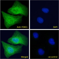
ARG63341 anti-FOXC1 antibody ICC/IF image
Immunofluorescence: Paraformaldehyde fixed U2OS cells permeabilized with 0.15% Triton. Cells were stained with ARG63341 anti-FOXC1 antibody (green) at 10 µg/ml dilution for 1 hour. DAPI (blue) for nuclear staining. Negative control: Unimmunized goat IgG (green) at 10 µg/ml dilution.
-
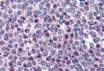
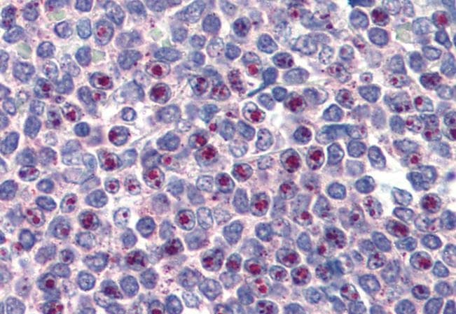
ARG63341 anti-FOXC1 antibody IHC-P image
Immunohistochemistry: Paraffin-embedded Human spleen tissue. Antigen Retrieval: Steam tissue section in Citrate buffer (pH 6.0). The tissue section was stained with ARG63341 anti-FOXC1 antibody at 3.75 µg/ml dilution followed by AP-staining.
-
ARG57983 anti-CDKN2A / p16INK4a antibody IHC-P image
Immunohistochemistry: Paraffin-embedded Human gastric stained with ARG57983 anti-CDKN2A / p16INK4a antibody.
-
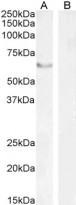
ARG63332 anti-FOXC2 antibody WB image
Western blot: 35 µg of HEK293 nucleus (A) and Human pancreas (B, negative control) lysates (in RIPA buffer) stained with ARG63332 anti-FOXC2 antibody at 2 µg/ml (A) and 1 µg/ml (B) dilutions and incubated at RT for 1 hour.
-
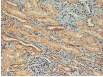
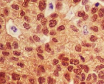
ARG63341 anti-FOXC1 antibody IHC-P image
Immunohistochemistry: paraffin embedded Human Kidney. (Steamed antigen retrieval with citrate buffer pH 6) stained with ARG63341 anti-FOXC1 antibody at 4 µg/ml dilution followed by HRP-staining.
-
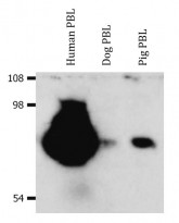
ARG62852 anti-CD44 antibody [MEM-263] WB image
Western blot: Isolated peripheral blood lymphocytes (PBL) of various species. Human PBL, Dog PBL and Pig PBL lysates stained with ARG62852 anti-CD44 antibody [MEM-263], in non-reducing conditions.
-
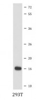
ARG57983 anti-CDKN2A / p16INK4a antibody WB image
Western blot: 293T cell lysate stained with ARG57983 anti-CDKN2A / p16INK4a antibody.
-
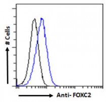
ARG63332 anti-FOXC2 antibody FACS image
Flow Cytometry: Paraformaldehyde-fixed HEK293 cells permeabilized with 0.5% Triton. Cells were stained with ARG63332 anti-FOXC2 antibody (blue line) at 10 µg/ml dilution for 1 hour, followed by incubation with Alexa FluorR 488 labelled secondary antibody. IgG control: Unimmunized goat IgG (black line).
-
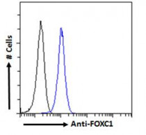
ARG63341 anti-FOXC1 antibody FACS image
Flow Cytometry: Paraformaldehyde-fixed HEK293 cells permeabilized with 0.5% Triton. Cells were stained with ARG63341 anti-FOXC1 antibody (blue line) at 10 µg/ml dilution for 1 hour, followed by incubation with Alexa FluorR 488 labelled secondary antibody. IgG control: Unimmunized goat IgG (black line).
-
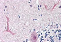
ARG63341 anti-FOXC1 antibody IHC-P image
Immunohistochemistry: Paraffin-embedded Human cerebellum tissue. Antigen Retrieval: Steam tissue section in Citrate buffer (pH 6.0). The tissue section was stained with ARG63341 anti-FOXC1 antibody at 3.75 µg/ml dilution followed by AP-staining.
-
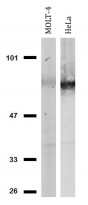
ARG62852 anti-CD44 antibody [MEM-263] WB image
Western blot: MOLT-4 and HeLa cell lysates stained with ARG62852 anti-CD44 antibody [MEM-263], in non-reducing conditions.


Specific References
KDF1 Promoted Proliferation, Migration and Invasion of Lung Adenocarcinoma Cells through Activating STAT3 and AKT Pathway
ARG65350: WB /


Expression and clinical significance of FOXC2 and E-cad in colon cancer tissues (FOXC2和E-cad在结肠癌组织中的表达及临床意义)
ARG63332: IHC-P / Human


