ARG62852
anti-CD44 antibody [MEM-263]
anti-CD44 antibody [MEM-263] for Flow cytometry,IHC-Formalin-fixed paraffin-embedded sections,Immunoprecipitation,Western blot and Human,Dog,Pig
Cancer antibody; Developmental Biology antibody; Immune System antibody; Chondrogenesis Study antibody
Overview
| Product Description | Mouse Monoclonal antibody [MEM-263] recognizes CD44 |
|---|---|
| Tested Reactivity | Hu, Dog, Pig |
| Predict Reactivity | Mk |
| Tested Application | FACS, IHC-P, IP, WB |
| Specificity | The clone MEM-263 reacts with extracellular (N-terminal) domain of standard CD44 (Phagocyte glycoprotein 1), a 80-95 kDa transmembrane glycoprotein (hyaladherin family) present on the most of cells and tissues (leukocytes, endothelial cells, mesenchymal cells, etc.); it is negative on platelets and hepatocytes. HLDA III; WS Code T 155 |
| Host | Mouse |
| Clonality | Monoclonal |
| Clone | MEM-263 |
| Isotype | IgG1 |
| Target Name | CD44 |
| Immunogen | COS-7 cells (African Green Monkey). |
| Conjugation | Un-conjugated |
| Alternate Names | MDU2; MDU3; GP90 lymphocyte homing/adhesion receptor; Hermes antigen; Extracellular matrix receptor III; PGP-I; Epican; CDW44; Phagocytic glycoprotein 1; Pgp1; HUTCH-I; MC56; Hyaluronate receptor; CD antigen CD44; Heparan sulfate proteoglycan; CD44 antigen; LHR; IN; HCELL; Phagocytic glycoprotein I; PGP-1; CSPG8; MIC4; ECMR-III; CDw44 |
Application Instructions
| Application Suggestion |
|
||||||||||
|---|---|---|---|---|---|---|---|---|---|---|---|
| Application Note | WB: 2 μg/ml dilution, 60 min on vertical incubator Positive control: Kg-1a human acute leukemia cell lysate JURKAT human leukemia T-cell lysate Sample preparation: Resuspend approx. 50 mil. cells in 1 ml cold Lysis buffer (1% laurylmaltoside in 20 mM Tris/Cl, 100 mM NaCl pH 8.2, 50 mM NaF including Protease inhibitor Cocktail). Incubate 60 min on ice. Centrifuge to remove cell debris. Mix lysate with non-reducing SDS-PAGE sample buffer. Application note: Non-reducing conditions. SDS-PAGE (6% separating gel). IHC-P: Positive tissue: uterus, myometrium * The dilutions indicate recommended starting dilutions and the optimal dilutions or concentrations should be determined by the scientist. |
Properties
| Form | Liquid |
|---|---|
| Purification | Purified from ascites by protein-A affinity chromatography. |
| Purity | > 95% (by SDS-PAGE) |
| Buffer | PBS (pH 7.4) and 15 mM Sodium azide |
| Preservative | 15 mM Sodium azide |
| Concentration | 1 mg/ml |
| Storage Instruction | For continuous use, store undiluted antibody at 2-8°C for up to a week. For long-term storage, aliquot and store at -20°C or below. Storage in frost free freezers is not recommended. Avoid repeated freeze/thaw cycles. Suggest spin the vial prior to opening. The antibody solution should be gently mixed before use. |
| Note | For laboratory research only, not for drug, diagnostic or other use. |
Bioinformation
| Database Links | |
|---|---|
| Background | CD44 is a transmembrane glycoprotein expressed on the surface of most cells, which serves as a receptor for hyaluronan. CD44 mediates angiogenesis, cell adhesion, proliferation and migration, it is thus important for lymphocyte activation, recirculation and homing, it can thus serve e.g. as a modulator of macrophage recruitment in response to pathogen. Although CD44 functions are essential for physiological activities of normal cells, elevated CD44 expression correlates with poor prognosis in many carcinomas, facilitating tumour growth and metastasis, antiapoptosis and directional motility of cancer cells. |
| Highlight | Related Antibody Duos and Panels: ARG30314 Chondrogenesis Marker Antibody Panel Related products: CD44 antibodies; CD44 ELISA Kits; CD44 Duos / Panels; Anti-Mouse IgG secondary antibodies; |
| Research Area | Cancer antibody; Developmental Biology antibody; Immune System antibody; Chondrogenesis Study antibody |
| Calculated MW | 82 kDa |
| PTM | Proteolytically cleaved in the extracellular matrix by specific proteinases (possibly MMPs) in several cell lines and tumors. N- and O-glycosylated. O-glycosylation contains more-or-less-sulfated chondroitin sulfate glycans, whose number may affect the accessibility of specific proteinases to their cleavage site(s). It is uncertain if O-glycosylation occurs on Thr-637 or Thr-638. Phosphorylated; activation of PKC results in the dephosphorylation of Ser-706 (constitutive phosphorylation site), and the phosphorylation of Ser-672. |
Images (2) Click the Picture to Zoom In
-
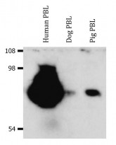
ARG62852 anti-CD44 antibody [MEM-263] WB image
Western blot: Isolated peripheral blood lymphocytes (PBL) of various species. Human PBL, Dog PBL and Pig PBL lysates stained with ARG62852 anti-CD44 antibody [MEM-263], in non-reducing conditions.
-
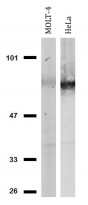
ARG62852 anti-CD44 antibody [MEM-263] WB image
Western blot: MOLT-4 and HeLa cell lysates stained with ARG62852 anti-CD44 antibody [MEM-263], in non-reducing conditions.


Clone References
Repeated autologous bone marrow-derived mesenchymal stem cell injections improve radiation-induced proctitis in pigs.


Loss of the malignant phenotype of human neuroblastoma cells by a catalytically inactive dominant-negative hTERT mutant.
Alternative pathway activation of complement by Shiga toxin promotes exuberant C3a formation that triggers microvascular thrombosis.
BL / Human


