ARG30318
CK7 / CK20 Carcinoma Antibody Duo
Component
| Cat No | Component Name | Host clonality | Reactivity | Application | Package |
|---|---|---|---|---|---|
| ARG66340 | anti-Cytokeratin 7 antibody [SQab1888] | Rabbit mAb | Hu, Ms | FACS, ICC/IF, IHC-P, IP, WB | 50 μl |
| ARG66248 | anti-Cytokeratin 20 antibody [SQab1737] | Rabbit mAb | Hu | FACS, ICC/IF, IHC-P, IP, WB | 50 μl |
Overview
| Product Description | CK7 and CK20 are the two most common cytokeratins used for identifying primary tumor sites. For example, CK7+/CK20+ is associated with carcinoma of transitional cell or ovarian origin; CK7+/CK20− is seen in many carcinomas including those of the lung, breast, thyroid, and female genital tract; CK7−/CK20+ is the hallmark of colorectal carcinoma and merkel cell carcinoma; and CK7−/CK20− is shown in carcinoma from prostate, hepatocellular, renal, or squamous cell origin. arigo's CK7/CK20 Carcinoma Antibody Duo comprises CK5 and CK20 antibodies. Both are rabbit monoclonal antibodies with excellent performance on IHC and other application. This antibody panel is an excellent solution for identifying primary tumor origin in metastatic cancer tissues. |
|---|---|
| Target Name | CK7 / CK20 Carcinoma |
| Alternate Names | CK7 / CK20 Carcinoma antibody; Cytokeratin 7 / Cytokeratin 20 Carcinoma antibody; Cytokeratin 20 antibody; Cytokeratin 7 antibody |
Properties
| Storage Instruction | For continuous use, store undiluted antibody at 2-8°C for up to a week. For long-term storage, aliquot and store at -20°C or below. Storage in frost free freezers is not recommended. Avoid repeated freeze/thaw cycles. Suggest spin the vial prior to opening. The antibody solution should be gently mixed before use. |
|---|---|
| Note | For laboratory research only, not for drug, diagnostic or other use. |
Bioinformation
| Gene Full Name | Cytokeratin 7 (CK7) / Cytokeratin 20 (CK20) Carcinoma Antibody Duo |
|---|---|
| Highlight | Related Product: anti-Cytokeratin 7 antibody; anti-Cytokeratin 20 antibody; |
Images (11) Click the Picture to Zoom In
-
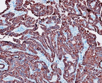
ARG66340 anti-Cytokeratin 7 antibody [SQab1888] IHC-P image
Immunohistochemistry: 10% Formalin-fixed and paraffin-embedded Mouse tumor tissue. Antigen Retrieval: Heat mediation was performed using Tris/EDTA buffer (pH 9.0). The tissue section was stained with ARG66340 anti-Cytokeratin 7 antibody [SQab1888] at 1:3000 dilution for 30 min at 37ºC.
-

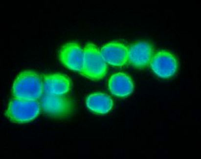
ARG66248 anti-Cytokeratin 20 antibody [SQab1737] ICC/IF image
Immunofluorescence: HT-29 cells were fixed with 4% paraformaldehyde for 30 min at RT, permeabilized with 0.1% Triton X-100 for 10 min at RT then blocked with 10% goat serum for half an hour at RT. Samples were stained with ARG66248 anti-Cytokeratin 20 antibody [SQab1737] (green) at 1:200, 4°C.
-
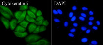
ARG66340 anti-Cytokeratin 7 antibody [SQab1888] ICC/IF image
Immunofluorescence: HeLa cells were fixed with 4% paraformaldehyde for 30 min at RT, permeabilized with 0.1% Triton X-100 for 10 min at RT then blocked with 10% goat serum for 30 min at RT. Cells were stained with ARG66340 anti-Cytokeratin 7 antibody [SQab1888] (green) at 1:2,000 and 4°C. DAPI (blue) was used as the nuclear counter stain.
-
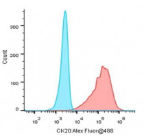
ARG66248 anti-Cytokeratin 20 antibody [SQab1737] FACS image
Flow Cytometry: HT-29 cells stained with ARG66248 anti-Cytokeratin 20 antibody [SQab1737] (Red). The cells were fixed with 4% paraformaldehyde (10 min) and then permeabilized with 0.1% TritonX-100 for 15 min. The cells were then incubated in the primary antibody at 1:1000 dilution in 1x PBS/1% BSA for 30 min at 4°C. The secondary antibody used was a Goat anti-rabbit Alexa Fluor® 488 (IgG H+L) at 1:2000 dilution for 20 min at 4°C. Unlabelled sample (Blue) was used as a control.
-
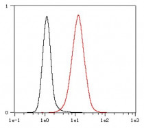
ARG66340 anti-Cytokeratin 7 antibody [SQab1888] FACS image
Flow Cytometry: HeLa cells were fixed with 4% paraformaldehyde (10 min) and then permeabilized with 0.1% TritonX-100 for 15 min. The cells were stained with ARG66340 anti-Cytokeratin 7 antibody [SQab1888] (red) at 1:800 dilution in 1x PBS/1% BSA for 30 min at RT, followed by Alexa Fluor® 488 labelled secondary antibody. Unlabelled sample (black) was used as a control.
-
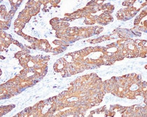
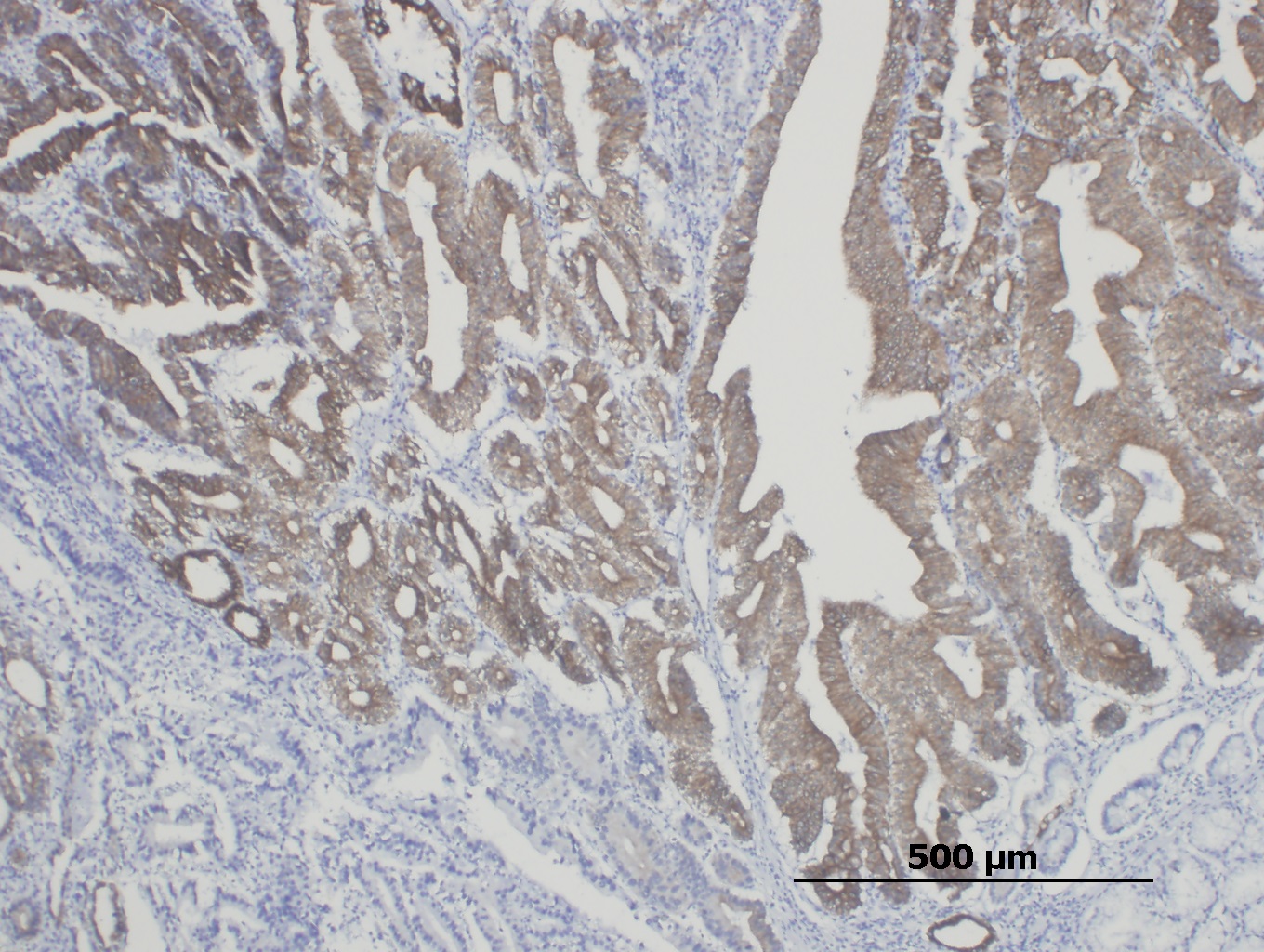
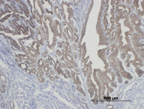
ARG66248 anti-Cytokeratin 20 antibody [SQab1737] IHC-P image
Immunohistochemistry: Formalin-fixed and paraffin-embedded colonic adenocarcinoma stained with ARG66248 anti-Cytokeratin 20 antibody [SQab1737] at 1:800. Antigen Retrieval: Boil tissue section in Tris/EDTA buffer (pH 9.0).
-
ARG66340 anti-Cytokeratin 7 antibody [SQab1888] IHC-P image
Immunohistochemistry: Formalin-fixed and paraffin-embedded Human thyroid cancer tissue. Antigen Retrieval: Heat mediation was performed using Tris/EDTA buffer (pH 9.0). The tissue section was stained with ARG66340 anti-Cytokeratin 7 antibody [SQab1888] at 1:3200 dilution.
-
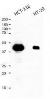
ARG66248 anti-Cytokeratin 20 antibody [SQab1737] WB image
Western blot: 10 μg of HCT-116 and HT-29 cell lysates stained with ARG66248 anti-Cytokeratin 20 antibody [SQab1737] at 1:5000 dilution.
-
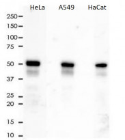
ARG66340 anti-Cytokeratin 7 antibody [SQab1888] WB image
Western blot: 10 µg of HeLa, A549 and HaCat cell lysates stained with ARG66340 anti-Cytokeratin 7 antibody [SQab1888] at 1:10000 dilution.
-
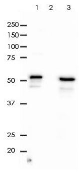
ARG66248 anti-Cytokeratin 20 antibody [SQab1737] IP image
Immunoprecipitation: Cytokeratin 20 was immunoprecipitated from 0.5 mg of HT-29 lysate with ARG66248 anti-Cytokeratin 20 antibody [SQab1737] at 1:50 dilution.
1. IP by using ARG66248 in HT-29 whole cell lysate
2. PBS instead of ARG66248 in HT-29 whole cell lysate
3. 10 µg of HT-29 whole cell lysate (input) -

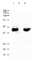
ARG66340 anti-Cytokeratin 7 antibody [SQab1888] IP image
Immunoprecipitation: 0.4 mg of HeLa whole cell lysate immunoprecipitated (1:50) and stained with ARG66340 anti-Cytokeratin 7 antibody [SQab1888]. 1) ARG66340 IP in HeLa whole cell lysate, 2) PBS instead of ARG66340 in HeLa whole cell lysate, and 3) HeLa whole cell lysate, 10 µg (input).