ARG30315
Brain Injury IHC Marker Antibody Duo (GFAP, MMP9)
Component
| Cat No | Component Name | Host clonality | Reactivity | Application | Package |
|---|---|---|---|---|---|
| ARG22191 | anti-MMP9 antibody [SB15c] | Mouse mAb | Hu | ELISA, ICC/IF, IHC-P, WB | 50 μg |
| ARG10122 | anti-GFAP antibody [GF5] | Mouse mAb | Hu, Ms, Rat | ELISA, ICC/IF, IHC-Fr, WB | 50 μg |
Overview
| Product Description | Glial fibrillary acidic protein (GFAP) is the major intermediate filament protein in mature astrocytes, a main type of glial cells in the central nervous system (CNS). GFAP is augmented in astrogliosis, a pathophysiology associated with degenerative and infectious/inflammatory brain disorders. Matrix metalloproteinase 9 (MMP-9) is a member of metalloproteinases family. It cleaves ECM and cell surface receptors allowing for synaptic and circuit level reorganization. MMP-9 level is increased after traumatic brain injury and involved in the following excitotoxic neuronal loss. arigo's ARG30292 Brain Injury IHC Marker Antibody Duo (GFAP, MMP9) comprise two antibodies against GFAP and MMP9, the pathological hallmark of CNS lesions, and is excellent for histological analysis of brain injury. Related news: Astrocyte-to-neuron conversion for Parkinson's disease treatment |
|---|---|
| Target Name | Brain Injury IHC Marker |
| Alternate Names | Brain Injury IHC Marker antibody; GFAP antibody; MMP9 antibody |
Properties
| Storage Instruction | For continuous use, store undiluted antibody at 2-8°C for up to a week. For long-term storage, aliquot and store at -20°C or below. Storage in frost free freezers is not recommended. Avoid repeated freeze/thaw cycles. Suggest spin the vial prior to opening. The antibody solution should be gently mixed before use. |
|---|---|
| Note | For laboratory research only, not for drug, diagnostic or other use. |
Bioinformation
| Gene Full Name | Antibody Duo for Brain Injury IHC Marker (GFAP, MMP9) |
|---|---|
| Highlight | Related Product: anti-MMP9 antibody; anti-GFAP antibody; |
Images (6) Click the Picture to Zoom In
-
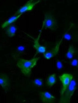
ARG10122 anti-GFAP antibody [GF5] ICC/IF image
Immunofluorescence: Rat astrocyte primary cell stained with ARG10122 anti-GFAP antibody [GF5] (green) at 1:200 dilution.
Cell nuclei was stained with DAPI (blue). -
ARG10122 anti-GFAP antibody [GF5] IHC-Fr image
Immunohistochemistry: Rat ventral horn of spinal cord stained with ARG10122 anti-GFAP antibody [GF5] at 1: 500 dilution.
From Chin-An Chen et al. Int J Med Sci. (2021), doi: 10.7150/ijms.65976, Fig. 4A.
-
ARG10122 anti-GFAP antibody [GF5] WB image
Western blot: 20 µg of Mouse brain and Rat brain lysates stained with ARG10122 anti-GFAP antibody [GF5] at 1:500 dilution.
-
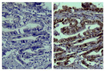
ARG22191 anti-MMP9 antibody [SB15c] IHC-P image
Immunohistochemistry: Paraffin-embedded Human gastric cancer tissue stained with ARG21961 Mouse IgG2a Isotype Control antibody [HOPC-1] (left) and ARG22191 anti-MMP9 antibody [SB15c] (right) followed by ARG23705 Goat anti-Mouse IgG2a (HRP) (pre-adsorbed), DAB and hematoxylin.
-
ARG10122 anti-GFAP antibody [GF5] WB image
Western blot: 30 µg of Rat brain lysate stained with ARG10122 anti-GFAP antibody [GF5] at 1:500 dilution.
Specific References


