ARG40315
anti-p70 S6 Kinase phospho (Thr389) antibody
anti-p70 S6 Kinase phospho (Thr389) antibody for ICC/IF,IHC-Formalin-fixed paraffin-embedded sections,Western blot and Mouse,Rat
Overview
| Product Description | Rabbit Polyclonal antibody recognizes p70 S6 Kinase phospho (Thr389) |
|---|---|
| Tested Reactivity | Ms, Rat |
| Tested Application | ICC/IF, IHC-P, WB |
| Host | Rabbit |
| Clonality | Polyclonal |
| Isotype | IgG |
| Target Name | p70 S6 Kinase |
| Antigen Species | Human |
| Immunogen | Phosphospecific peptide around Thr389 of Human p70 S6 Kinase. |
| Conjugation | Un-conjugated |
| Alternate Names | p70 S6 kinase alpha; p70(S6K)-alpha; p70-alpha; p70 S6KA; S6K; STK14A; S6K1; p70-S6K 1; Ribosomal protein S6 kinase I; 70 kDa ribosomal protein S6 kinase 1; S6K-beta-1; P70S6K1; PS6K; p70 ribosomal S6 kinase alpha; p70-S6K; EC 2.7.11.1; Serine/threonine-protein kinase 14A; p70 S6K-alpha; Ribosomal protein S6 kinase beta-1 |
Application Instructions
| Application Suggestion |
|
||||||||
|---|---|---|---|---|---|---|---|---|---|
| Application Note | * The dilutions indicate recommended starting dilutions and the optimal dilutions or concentrations should be determined by the scientist. | ||||||||
| Positive Control | NIH/3T3 | ||||||||
| Observed Size | 70 kDa |
Properties
| Form | Liquid |
|---|---|
| Purification | Affinity purified. |
| Buffer | PBS (pH 7.3), 0.02% Sodium azide and 50% Glycerol. |
| Preservative | 0.02% Sodium azide |
| Stabilizer | 50% Glycerol |
| Storage Instruction | For continuous use, store undiluted antibody at 2-8°C for up to a week. For long-term storage, aliquot and store at -20°C. Storage in frost free freezers is not recommended. Avoid repeated freeze/thaw cycles. Suggest spin the vial prior to opening. The antibody solution should be gently mixed before use. |
| Note | For laboratory research only, not for drug, diagnostic or other use. |
Bioinformation
| Database Links |
Swiss-port # P67999 Rat Ribosomal protein S6 kinase beta-1 Swiss-port # Q8BSK8 Mouse Ribosomal protein S6 kinase beta-1 |
|---|---|
| Gene Symbol | RPS6KB1 |
| Gene Full Name | ribosomal protein S6 kinase, 70kDa, polypeptide 1 |
| Background | This gene encodes a member of the ribosomal S6 kinase family of serine/threonine kinases. The encoded protein responds to mTOR (mammalian target of rapamycin) signaling to promote protein synthesis, cell growth, and cell proliferation. Activity of this gene has been associated with human cancer. Alternatively spliced transcript variants have been observed. The use of alternative translation start sites results in isoforms with longer or shorter N-termini which may differ in their subcellular localizations. There are two pseudogenes for this gene on chromosome 17. [provided by RefSeq, Jan 2013] |
| Function | Serine/threonine-protein kinase that acts downstream of mTOR signaling in response to growth factors and nutrients to promote cell proliferation, cell growth and cell cycle progression. Regulates protein synthesis through phosphorylation of EIF4B, RPS6 and EEF2K, and contributes to cell survival by repressing the pro-apoptotic function of BAD. Under conditions of nutrient depletion, the inactive form associates with the EIF3 translation initiation complex. Upon mitogenic stimulation, phosphorylation by the mammalian target of rapamycin complex 1 (mTORC1) leads to dissociation from the EIF3 complex and activation. The active form then phosphorylates and activates several substrates in the pre-initiation complex, including the EIF2B complex and the cap-binding complex component EIF4B. Also controls translation initiation by phosphorylating a negative regulator of EIF4A, PDCD4, targeting it for ubiquitination and subsequent proteolysis. Promotes initiation of the pioneer round of protein synthesis by phosphorylating POLDIP3/SKAR. In response to IGF1, activates translation elongation by phosphorylating EEF2 kinase (EEF2K), which leads to its inhibition and thus activation of EEF2. Also plays a role in feedback regulation of mTORC2 by mTORC1 by phosphorylating RICTOR, resulting in the inhibition of mTORC2 and AKT1 signaling. Mediates cell survival by phosphorylating the pro-apoptotic protein BAD and suppressing its pro-apoptotic function. Phosphorylates mitochondrial URI1 leading to dissociation of a URI1-PPP1CC complex. The free mitochondrial PPP1CC can then dephosphorylate RPS6KB1 at Thr-412, which is proposed to be a negative feedback mechanism for the RPS6KB1 anti-apoptotic function. Mediates TNF-alpha-induced insulin resistance by phosphorylating IRS1 at multiple serine residues, resulting in accelerated degradation of IRS1. In cells lacking functional TSC1-2 complex, constitutively phosphorylates and inhibits GSK3B. May be involved in cytoskeletal rearrangement through binding to neurabin. Phosphorylates and activates the pyrimidine biosynthesis enzyme CAD, downstream of MTOR. [UniProt] |
| Cellular Localization | Cell junction, synapse, synaptosome. Mitochondrion outer membrane. Mitochondrion. Note=Colocalizes with URI1 at mitochondrion. Isoform Alpha I: Nucleus. Cytoplasm. Isoform Alpha II: Cytoplasm. [UniProt] |
| Calculated MW | 59 kDa |
| PTM | Phosphorylation at Thr-412 is regulated by mTORC1. The phosphorylation at this site is maintained by an agonist-dependent autophosphorylation mechanism (By similarity). Activated by phosphorylation at Thr-252 by PDPK1. Dephosphorylation by PPP1CC at Thr-412 in mitochondrion. [UniProt] |
Images (3) Click the Picture to Zoom In
-
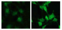
ARG40315 anti-p70 S6 Kinase phospho (Thr389) antibody ICC/IF image
Immunofluorescence: C6 cells stained with ARG40315 anti-p70 S6 Kinase phospho (Thr389) antibody at 1:100 dilution. Cells were untreated (serum starvation, left) or treated with serum (right) overnight at 37°C.
-
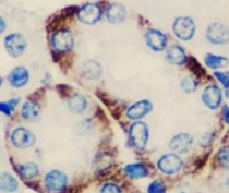
ARG40315 anti-p70 S6 Kinase phospho (Thr389) antibody IHC-P image
Immunohistochemistry: Paraffin-embedded Mouse kidney stained with ARG40315 anti-p70 S6 Kinase phospho (Thr389) antibody at 1:100 dilution.
-
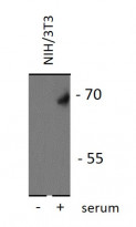
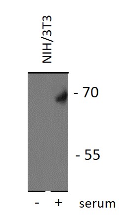
ARG40315 anti-p70 S6 Kinase phospho (Thr389) antibody WB image
Western blot: 25 µg of NIH/3T3 cell lysates stained with ARG40315 anti-p70 S6 Kinase phospho (Thr389) antibody at 1:2000 dilution. NIH/3T3 cells were starvation for overnight before treated with 10% FBS at 37°C for 30 minutes (right lane) or without FBS treatment as negative control (left lane).