ARG10654
anti-p63 antibody
anti-p63 antibody for Flow cytometry,ICC/IF,IHC-Formalin-fixed paraffin-embedded sections,Western blot and Human,Mouse,Rat
Overview
| Product Description | Rabbit Polyclonal antibody recognizes p63 |
|---|---|
| Tested Reactivity | Hu, Ms, Rat |
| Tested Application | FACS, ICC/IF, IHC-P, WB |
| Host | Rabbit |
| Clonality | Polyclonal |
| Isotype | IgG |
| Target Name | p63 |
| Antigen Species | Human |
| Immunogen | Synthetic peptide around aa. 82-98 of Human p63. (DMDARRNKQQRIKEEGE) |
| Conjugation | Un-conjugated |
| Alternate Names | p63; Tumor protein p73-like; B(p51A); AIS; p53CP; p73L; p73H; p40; EEC3; TP63; NBP; Chronic ulcerative stomatitis protein; TP53CP; CUSP; B(p51B); TP73L; p51; Transformation-related protein 63; Keratinocyte transcription factor KET; SHFM4; TP53L; RHS; LMS; Tumor protein 63; OFC8; KET |
Application Instructions
| Application Suggestion |
|
||||||||||
|---|---|---|---|---|---|---|---|---|---|---|---|
| Application Note | The antibody is not suitable for mouse and rat samples by IHC-P application (might get week signal). * The dilutions indicate recommended starting dilutions and the optimal dilutions or concentrations should be determined by the scientist. |
Properties
| Form | Liquid |
|---|---|
| Purification | Affinity purification with immunogen. |
| Buffer | PBS, 0.025% Sodium azide and 2.5% BSA. |
| Preservative | 0.025% Sodium azide |
| Stabilizer | 2.5% BSA |
| Concentration | 0.5 mg/ml |
| Storage Instruction | For continuous use, store undiluted antibody at 2-8°C for up to a week. For long-term storage, aliquot and store at -20°C or below. Storage in frost free freezers is not recommended. Avoid repeated freeze/thaw cycles. Suggest spin the vial prior to opening. The antibody solution should be gently mixed before use. |
| Note | For laboratory research only, not for drug, diagnostic or other use. |
Bioinformation
| Database Links | |
|---|---|
| Gene Symbol | TP63 |
| Gene Full Name | tumor protein p63 |
| Background | This gene encodes a member of the p53 family of transcription factors. An animal model, p63 -/- mice, has been useful in defining the role this protein plays in the development and maintenance of stratified epithelial tissues. p63 -/- mice have several developmental defects which include the lack of limbs and other tissues, such as teeth and mammary glands, which develop as a result of interactions between mesenchyme and epithelium. Mutations in this gene are associated with ectodermal dysplasia, and cleft lip/palate syndrome 3 (EEC3); split-hand/foot malformation 4 (SHFM4); ankyloblepharon-ectodermal defects-cleft lip/palate; ADULT syndrome (acro-dermato-ungual-lacrimal-tooth); limb-mammary syndrome; Rap-Hodgkin syndrome (RHS); and orofacial cleft 8. Both alternative splicing and the use of alternative promoters results in multiple transcript variants encoding different proteins. Many transcripts encoding different proteins have been reported but the biological validity and the full-length nature of these variants have not been determined. [provided by RefSeq, Jul 2008] |
| Function | Acts as a sequence specific DNA binding transcriptional activator or repressor. The isoforms contain a varying set of transactivation and auto-regulating transactivation inhibiting domains thus showing an isoform specific activity. Isoform 2 activates RIPK4 transcription. May be required in conjunction with TP73/p73 for initiation of p53/TP53 dependent apoptosis in response to genotoxic insults and the presence of activated oncogenes. Involved in Notch signaling by probably inducing JAG1 and JAG2. Plays a role in the regulation of epithelial morphogenesis. The ratio of DeltaN-type and TA*-type isoforms may govern the maintenance of epithelial stem cell compartments and regulate the initiation of epithelial stratification from the undifferentiated embryonal ectoderm. Required for limb formation from the apical ectodermal ridge. Activates transcription of the p21 promoter. [UniProt] |
| Calculated MW | 77 kDa |
| PTM | May be sumoylated. Ubiquitinated. Polyubiquitination involves WWP1 and leads to proteasomal degradation of this protein. |
Images (8) Click the Picture to Zoom In
-
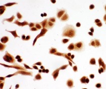
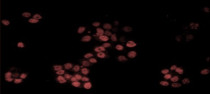
ARG10654 anti-p63 antibody ICC/IF image
Immunocytochemistry: A549 cells stained with ARG10654 anti-p63 antibody.
-
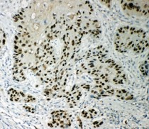
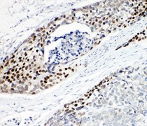
ARG10654 anti-p63 antibody IHC-P image
Immunohistochemistry: Paraffin-embedded Human esophageal squamous cell carcinoma tissue stained with ARG10654 anti-p63 antibody.
-
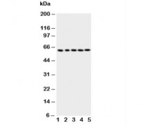
ARG10654 anti-p63 antibody WB image
Western blot: 1) HeLa, 2) SMMC-7721, 3) COLO320, 4) A549, and 5) SGC-7901 cell lysates stained with ARG10654 anti-p63 antibody.
-
ARG10654 anti-p63 antibody ICC/IF image
Immunofluorescence: A431 stained with ARG10654 anti-p63 antibody at 1:100
-
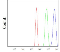
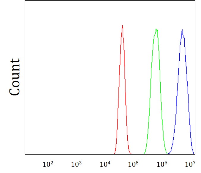
ARG10654 anti-p63 antibody FACS image
Flow Cytometry: A431 cells were stained with ARG10654 anti-p63 antibody (red) at 1:500 dilution/10^6 cells.
-
ARG10654 anti-p63 antibody IHC-P image
Immunohistochemistry: Paraffin-embedded Human esophageal squamous cell carcinoma tissue stained with ARG10654 anti-p63 antibody.
-
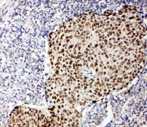
ARG10654 anti-p63 antibody IHC-P image
Immunohistochemistry: Paraffin-embedded Human lung cancer tissue stained with ARG10654 anti-p63 antibody.
-
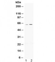
ARG10654 anti-p63 antibody WB image
Western blot: 50 µg of 1) Rat heart and 2) Mouse thymus lysates stained with ARG10654 anti-p63 antibody at 0.5 µg/ml dilution.