ARG51833
anti-c-Cbl phospho (Tyr700) antibody
anti-c-Cbl phospho (Tyr700) antibody for ICC/IF,IHC-Formalin-fixed paraffin-embedded sections,Western blot and Human
Cancer antibody; Cell Biology and Cellular Response antibody; Gene Regulation antibody; Signaling Transduction antibody
Overview
| Product Description | Rabbit Polyclonal antibody recognizes c-Cbl phospho (Tyr700) |
|---|---|
| Tested Reactivity | Hu |
| Tested Application | ICC/IF, IHC-P, WB |
| Host | Rabbit |
| Clonality | Polyclonal |
| Isotype | IgG |
| Target Name | c-Cbl |
| Antigen Species | Human |
| Immunogen | Peptide sequence around phosphorylation site of tyrosine 700 (T-E-Y(p)-M-T) derived from Human c-Cbl. |
| Conjugation | Un-conjugated |
| Alternate Names | Signal transduction protein CBL; C-CBL; EC 6.3.2.-; FRA11B; Casitas B-lineage lymphoma proto-oncogene; Proto-oncogene c-Cbl; RNF55; CBL2; E3 ubiquitin-protein ligase CBL; RING finger protein 55; NSLL |
Application Instructions
| Application Suggestion |
|
||||||||
|---|---|---|---|---|---|---|---|---|---|
| Application Note | * The dilutions indicate recommended starting dilutions and the optimal dilutions or concentrations should be determined by the scientist. |
Properties
| Form | Liquid |
|---|---|
| Purification | Antibodies were produced by immunizing rabbits with KLH-conjugated synthetic phosphopeptide. Antibodies were purified by affinity-chromatography using epitope-specific phosphopeptide. In addition, non-phospho specific antibodies were removed by chromatogramphy using non-phosphopeptide. |
| Buffer | PBS (without Mg2+ and Ca2+, pH 7.4), 150mM NaCl, 0.02% Sodium azide and 50% Glycerol. |
| Preservative | 0.02% Sodium azide |
| Stabilizer | 50% Glycerol |
| Concentration | 1 mg/ml |
| Storage Instruction | For continuous use, store undiluted antibody at 2-8°C for up to a week. For long-term storage, aliquot and store at -20°C. Storage in frost free freezers is not recommended. Avoid repeated freeze/thaw cycles. Suggest spin the vial prior to opening. The antibody solution should be gently mixed before use. |
| Note | For laboratory research only, not for drug, diagnostic or other use. |
Bioinformation
| Database Links | |
|---|---|
| Gene Symbol | CBL |
| Gene Full Name | Cbl proto-oncogene, E3 ubiquitin protein ligase |
| Background | Participates in signal transduction in hematopoietic cells. Adapter protein that functions as a negative regulator of many signaling pathways that start from receptors at the cell surface. Acts as an E3 ubiquitin-protein ligase, which accepts ubiquitin from specific E2 ubiquitin-conjugating enzymes, and then transfers it to substrates promoting their degradation by the proteasome. Recognizes activated receptor tyrosine kinases, including PDGFA, EGF and CSF1, and terminates signaling. |
| Function | Adapter protein that functions as a negative regulator of many signaling pathways that are triggered by activation of cell surface receptors. Acts as an E3 ubiquitin-protein ligase, which accepts ubiquitin from specific E2 ubiquitin-conjugating enzymes, and then transfers it to substrates promoting their degradation by the proteasome. Recognizes activated receptor tyrosine kinases, including KIT, FLT1, FGFR1, FGFR2, PDGFRA, PDGFRB, EGFR, CSF1R, EPHA8 and KDR and terminates signaling. Recognizes membrane-bound HCK, SRC and other kinases of the SRC family and mediates their ubiquitination and degradation. Participates in signal transduction in hematopoietic cells. Plays an important role in the regulation of osteoblast differentiation and apoptosis. Essential for osteoclastic bone resorption. The 'Tyr-731' phosphorylated form induces the activation and recruitment of phosphatidylinositol 3-kinase to the cell membrane in a signaling pathway that is critical for osteoclast function. May be functionally coupled with the E2 ubiquitin-protein ligase UB2D3. [UniProt] |
| Research Area | Cancer antibody; Cell Biology and Cellular Response antibody; Gene Regulation antibody; Signaling Transduction antibody |
| Calculated MW | 100 kDa |
| PTM | Phosphorylated on tyrosine residues by ALK, EGFR, SYK, FYN and ZAP70 (By similarity). Phosphorylated on tyrosine residues in response to FLT1 and KIT signaling. Phosphorylated on tyrosine residues by INSR and FGR. Phosphorylated on several tyrosine residues by constitutively activated FGFR3. Not phosphorylated at Tyr-731 by FGFR3. Phosphorylated on tyrosine residues by activated CSF1R, PDGFRA and PDGFRB. Phosphorylated on tyrosine residues by HCK. Ubiquitinated, leading to its degradation via the proteasome. |
Images (3) Click the Picture to Zoom In
-

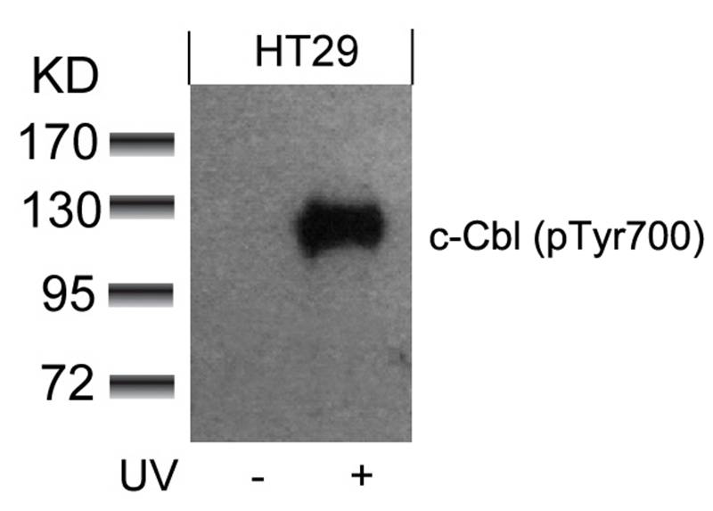
ARG51833 anti-c-Cbl phospho (Tyr700) antibody WB image
Western blot: Extracts from HT29 cells untreated or treated with UV stained with ARG51833 anti-c-Cbl phospho (Tyr700) antibody.
-
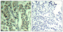
ARG51833 anti-c-Cbl phospho (Tyr700) antibody IHC-P image
Immunohistochemistry: Paraffin-embedded Human breast carcinoma tissue stained with ARG51833 anti-c-Cbl phospho (Tyr700) antibody (left) or the same antibody preincubated with blocking peptide (right).
-
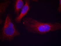
ARG51833 anti-c-Cbl phospho (Tyr700) antibody ICC/IF image
Immunofluorescence: methanol-fixed HeLa cells stained with ARG51833 anti-c-Cbl phospho (Tyr700) antibody.