ARG63177
anti-c-Cbl antibody
anti-c-Cbl antibody for Western blot and Mouse
Cancer antibody; Cell Biology and Cellular Response antibody; Gene Regulation antibody; Signaling Transduction antibody
Overview
| Product Description | Goat Polyclonal antibody recognizes c-Cbl |
|---|---|
| Tested Reactivity | Ms |
| Predict Reactivity | Hu, Rat, Cow, Dog, Pig |
| Tested Application | WB |
| Host | Goat |
| Clonality | Polyclonal |
| Isotype | IgG |
| Target Name | c-Cbl |
| Antigen Species | Human |
| Immunogen | C-REFVSISSPAHVAT |
| Conjugation | Un-conjugated |
| Alternate Names | Signal transduction protein CBL; C-CBL; EC 6.3.2.-; FRA11B; Casitas B-lineage lymphoma proto-oncogene; Proto-oncogene c-Cbl; RNF55; CBL2; E3 ubiquitin-protein ligase CBL; RING finger protein 55; NSLL |
Application Instructions
| Application Suggestion |
|
||||
|---|---|---|---|---|---|
| Application Note | WB: Recommend incubate at RT for 1h. * The dilutions indicate recommended starting dilutions and the optimal dilutions or concentrations should be determined by the scientist. |
Properties
| Form | Liquid |
|---|---|
| Purification | Purified from goat serum by antigen affinity chromatography. |
| Buffer | Tris saline (pH 7.3), 0.02% Sodium azide and 0.5% BSA. |
| Preservative | 0.02% Sodium azide |
| Stabilizer | 0.5% BSA |
| Concentration | 0.5 mg/ml |
| Storage Instruction | For continuous use, store undiluted antibody at 2-8°C for up to a week. For long-term storage, aliquot and store at -20°C or below. Storage in frost free freezers is not recommended. Avoid repeated freeze/thaw cycles. Suggest spin the vial prior to opening. The antibody solution should be gently mixed before use. |
| Note | For laboratory research only, not for drug, diagnostic or other use. |
Bioinformation
| Database Links | |
|---|---|
| Background | This gene is a proto-oncogene that encodes a RING finger E3 ubiquitin ligase. The encoded protein is one of the enzymes required for targeting substrates for degradation by the proteasome. This protein mediates the transfer of ubiquitin from ubiquitin conjugating enzymes (E2) to specific substrates. This protein also contains an N-terminal phosphotyrosine binding domain that allows it to interact with numerous tyrosine-phosphorylated substrates and target them for proteasome degradation. As such it functions as a negative regulator of many signal transduction pathways. This gene has been found to be mutated or translocated in many cancers including acute myeloid leukaemia. Mutations in this gene are also the cause of Noonan syndrome-like disorder. [provided by RefSeq, Mar 2012] |
| Research Area | Cancer antibody; Cell Biology and Cellular Response antibody; Gene Regulation antibody; Signaling Transduction antibody |
| Calculated MW | 100 kDa |
| PTM | Phosphorylated on tyrosine residues by ALK, EGFR, SYK, FYN and ZAP70 (By similarity). Phosphorylated on tyrosine residues in response to FLT1 and KIT signaling. Phosphorylated on tyrosine residues by INSR and FGR. Phosphorylated on several tyrosine residues by constitutively activated FGFR3. Not phosphorylated at Tyr-731 by FGFR3. Phosphorylated on tyrosine residues by activated CSF1R, PDGFRA and PDGFRB. Phosphorylated on tyrosine residues by HCK. Ubiquitinated, leading to its degradation via the proteasome. |
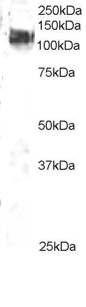
Images (1) Click the Picture to Zoom In
-

ARG63177 anti-c-Cbl antibody WB image
Western blot: 3T3 lysate (RIPA buffer, 35 µg total protein per lane) stained with ARG63177 anti-c-Cbl antibody at 2 µg/ml dilution.