ARG58066
anti-c Abl antibody
anti-c Abl antibody for ICC/IF,IHC-Formalin-fixed paraffin-embedded sections,Western blot and Human,Mouse,Rat
Overview
| Product Description | Rabbit Polyclonal antibody recognizes c Abl |
|---|---|
| Tested Reactivity | Hu, Ms, Rat |
| Tested Application | ICC/IF, IHC-P, WB |
| Host | Rabbit |
| Clonality | Polyclonal |
| Isotype | IgG |
| Target Name | c Abl |
| Antigen Species | Human |
| Immunogen | Synthetic peptide of Human c-Abl. |
| Conjugation | Un-conjugated |
| Alternate Names | bcr/abl; Proto-oncogene c-Abl; c-ABL; Abelson murine leukemia viral oncogene homolog 1; ABL; Abelson tyrosine-protein kinase 1; JTK7; Tyrosine-protein kinase ABL1; p150; v-abl; c-ABL1; EC 2.7.10.2 |
Application Instructions
| Application Suggestion |
|
||||||||
|---|---|---|---|---|---|---|---|---|---|
| Application Note | * The dilutions indicate recommended starting dilutions and the optimal dilutions or concentrations should be determined by the scientist. | ||||||||
| Positive Control | HeLa | ||||||||
| Observed Size | ~ 140 kDa |
Properties
| Form | Liquid |
|---|---|
| Purification | Affinity purified. |
| Buffer | PBS (pH 7.3), 0.02% Sodium azide and 50% Glycerol. |
| Preservative | 0.02% Sodium azide |
| Stabilizer | 50% Glycerol |
| Storage Instruction | For continuous use, store undiluted antibody at 2-8°C for up to a week. For long-term storage, aliquot and store at -20°C. Storage in frost free freezers is not recommended. Avoid repeated freeze/thaw cycles. Suggest spin the vial prior to opening. The antibody solution should be gently mixed before use. |
| Note | For laboratory research only, not for drug, diagnostic or other use. |
Bioinformation
| Database Links | |
|---|---|
| Gene Symbol | ABL1 |
| Gene Full Name | ABL proto-oncogene 1, non-receptor tyrosine kinase |
| Background | This gene is a protooncogene that encodes a protein tyrosine kinase involved in a variety of cellular processes, including cell division, adhesion, differentiation, and response to stress. The activity of the protein is negatively regulated by its SH3 domain, whereby deletion of the region encoding this domain results in an oncogene. The ubiquitously expressed protein has DNA-binding activity that is regulated by CDC2-mediated phosphorylation, suggesting a cell cycle function. This gene has been found fused to a variety of translocation partner genes in various leukemias, most notably the t(9;22) translocation that results in a fusion with the 5' end of the breakpoint cluster region gene (BCR; MIM:151410). Alternative splicing of this gene results in two transcript variants, which contain alternative first exons that are spliced to the remaining common exons. [provided by RefSeq, Aug 2014] |
| Function | Non-receptor tyrosine-protein kinase that plays a role in many key processes linked to cell growth and survival such as cytoskeleton remodeling in response to extracellular stimuli, cell motility and adhesion, receptor endocytosis, autophagy, DNA damage response and apoptosis. Coordinates actin remodeling through tyrosine phosphorylation of proteins controlling cytoskeleton dynamics like WASF3 (involved in branch formation); ANXA1 (involved in membrane anchoring); DBN1, DBNL, CTTN, RAPH1 and ENAH (involved in signaling); or MAPT and PXN (microtubule-binding proteins). Phosphorylation of WASF3 is critical for the stimulation of lamellipodia formation and cell migration. Involved in the regulation of cell adhesion and motility through phosphorylation of key regulators of these processes such as BCAR1, CRK, CRKL, DOK1, EFS or NEDD9. Phosphorylates multiple receptor tyrosine kinases and more particularly promotes endocytosis of EGFR, facilitates the formation of neuromuscular synapses through MUSK, inhibits PDGFRB-mediated chemotaxis and modulates the endocytosis of activated B-cell receptor complexes. Other substrates which are involved in endocytosis regulation are the caveolin (CAV1) and RIN1. Moreover, ABL1 regulates the CBL family of ubiquitin ligases that drive receptor down-regulation and actin remodeling. Phosphorylation of CBL leads to increased EGFR stability. Involved in late-stage autophagy by regulating positively the trafficking and function of lysosomal components. ABL1 targets to mitochondria in response to oxidative stress and thereby mediates mitochondrial dysfunction and cell death. ABL1 is also translocated in the nucleus where it has DNA-binding activity and is involved in DNA-damage response and apoptosis. Many substrates are known mediators of DNA repair: DDB1, DDB2, ERCC3, ERCC6, RAD9A, RAD51, RAD52 or WRN. Activates the proapoptotic pathway when the DNA damage is too severe to be repaired. Phosphorylates TP73, a primary regulator for this type of damage-induced apoptosis. Phosphorylates the caspase CASP9 on 'Tyr-153' and regulates its processing in the apoptotic response to DNA damage. Phosphorylates PSMA7 that leads to an inhibition of proteasomal activity and cell cycle transition blocks. ABL1 acts also as a regulator of multiple pathological signaling cascades during infection. Several known tyrosine-phosphorylated microbial proteins have been identified as ABL1 substrates. This is the case of A36R of Vaccinia virus, Tir (translocated intimin receptor) of pathogenic E.coli and possibly Citrobacter, CagA (cytotoxin-associated gene A) of H.pylori, or AnkA (ankyrin repeat-containing protein A) of A.phagocytophilum. Pathogens can highjack ABL1 kinase signaling to reorganize the host actin cytoskeleton for multiple purposes, like facilitating intracellular movement and host cell exit. Finally, functions as its own regulator through autocatalytic activity as well as through phosphorylation of its inhibitor, ABI1. [UniProt] |
| Cellular Localization | Cytoplasm, cytoskeleton, Nucleus, Mitochondrion, Nucleus membrane, Lipid-anchor. [UniProt] |
| Calculated MW | 123 kDa |
| PTM | Acetylated at Lys-711 by EP300 which promotes the cytoplasmic translocation. Phosphorylation at Tyr-70 by members of the SRC family of kinases disrupts SH3 domain-based autoinhibitory interactions and intermolecular associations, such as that with ABI1, and also enhances kinase activity. Phosphorylation at Tyr-226 and Tyr-393 correlate with increased activity. DNA damage-induced activation of ABL1 requires the function of ATM and Ser-446 phosphorylation (By similarity). Phosphorylation at Ser-569 has been attributed to a CDC2-associated kinase and is coupled to cell division (By similarity). Phosphorylation at Ser-618 and Ser-619 by PAK2 increases binding to CRK and reduces binding to ABI1. Phosphorylation on Thr-735 is required for binding 14-3-3 proteins for cytoplasmic translocation. Phosphorylated by PRKDC (By similarity). Polyubiquitinated. Polyubiquitination of ABL1 leads to degradation. [UniProt] |
Images (2) Click the Picture to Zoom In
-
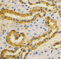
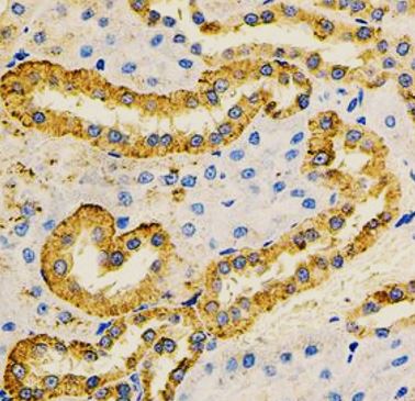
ARG58066 anti-c Abl antibody IHC-P image
Immunohistochemistry: Paraffin-embedded Rat kidney stained with ARG58066 anti-c Abl antibody at 1:100 dilution.
-
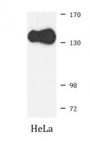
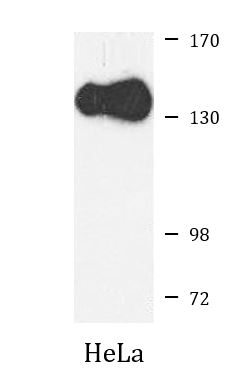
ARG58066 anti-c Abl antibody WB image
Western blot: 25 µg of HeLa cell lysate stained with ARG58066 anti-c Abl antibody at 1:1000 dilution.