ARG66170
anti-beta Tubulin antibody
anti-beta Tubulin antibody for IHC-Formalin-fixed paraffin-embedded sections,Western blot and Human,Mouse,Rat
Overview
| Product Description | Mouse Monoclonal antibody recognizes beta Tubulin |
|---|---|
| Tested Reactivity | Hu, Ms, Rat |
| Tested Application | IHC-P, WB |
| Specificity | The antibody detects endogenous beta Tubulin proteins. |
| Host | Mouse |
| Clonality | Monoclonal |
| Target Name | beta Tubulin |
| Antigen Species | Human |
| Immunogen | Synthetic peptide of Human beta Tubulin. |
| Conjugation | Un-conjugated |
| Alternate Names | Tubulin beta-1 chain |
Application Instructions
| Application Suggestion |
|
||||||
|---|---|---|---|---|---|---|---|
| Application Note | IHC-P: Antigen Retrieval: Boil tissue section in Sodium citrate buffer (pH 6.0) for 20 min. * The dilutions indicate recommended starting dilutions and the optimal dilutions or concentrations should be determined by the scientist. |
Properties
| Form | Liquid |
|---|---|
| Purification | Affinity purification with immunogen. |
| Buffer | PBS (pH 7.4), 0.02% Sodium azide and 50% Glycerol. |
| Preservative | 0.02% Sodium azide |
| Stabilizer | 50% Glycerol |
| Concentration | 1 mg/ml |
| Storage Instruction | For continuous use, store undiluted antibody at 2-8°C for up to a week. For long-term storage, aliquot and store at -20°C. Storage in frost free freezers is not recommended. Avoid repeated freeze/thaw cycles. Suggest spin the vial prior to opening. The antibody solution should be gently mixed before use. |
| Note | For laboratory research only, not for drug, diagnostic or other use. |
Bioinformation
| Database Links | |
|---|---|
| Gene Symbol | TUBB1 |
| Gene Full Name | tubulin, beta 1 class VI |
| Background | Beta tubulins are one of two core protein families (alpha and beta tubulins) that heterodimerize and assemble to form microtubules. This protein is specifically expressed in platelets and megakaryocytes and may be involved in proplatelet production and platelet release. A mutations in this gene is associated with autosomal dominant macrothrombocytopenia. Two pseudogenes of this gene are found on chromosome Y. [provided by RefSeq, Jul 2010] |
| Function | Tubulins is the major constituent of microtubules. It binds two moles of GTP, one at an exchangeable site on the beta chain and one at a non-exchangeable site on the alpha chain. [UniProt] |
| Calculated MW | 50 kDa |
| PTM | Some glutamate residues at the C-terminus are polyglutamylated, resulting in polyglutamate chains on the gamma-carboxyl group (PubMed:26875866). Polyglutamylation plays a key role in microtubule severing by spastin (SPAST). SPAST preferentially recognizes and acts on microtubules decorated with short polyglutamate tails: severing activity by SPAST increases as the number of glutamates per tubulin rises from one to eight, but decreases beyond this glutamylation threshold (PubMed:26875866). Some glutamate residues at the C-terminus are monoglycylated but not polyglycylated due to the absence of functional TTLL10 in human. Monoglycylation is mainly limited to tubulin incorporated into axonemes (cilia and flagella). Both polyglutamylation and monoglycylation can coexist on the same protein on adjacent residues, and lowering glycylation levels increases polyglutamylation, and reciprocally. The precise function of monoglycylation is still unclear (Probable). Phosphorylated on Ser-172 by CDK1 during the cell cycle, from metaphase to telophase, but not in interphase. This phosphorylation inhibits tubulin incorporation into microtubules. |
Images (16) Click the Picture to Zoom In
-
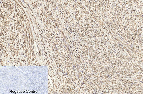
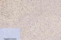
ARG66170 anti-beta Tubulin antibody IHC-P image
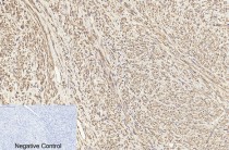
Immunohistochemistry: Paraffin-embedded Human uterus cancer tissue stained with ARG66170 anti-beta Tubulin antibody at 1:200 dilution (4°C, overnight). Antigen Retrieval: Boil tissue section in Sodium citrate buffer (pH 6.0) for 20 min.
Negative control was used by secondary antibody only.
-
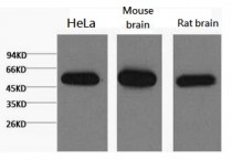
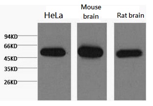
ARG66170 anti-beta Tubulin antibody WB image
Western blot: 1) HeLa, 2) Mouse brain, and 3) Rat brain lysates stained with ARG66170 anti-beta Tubulin antibody at 1:5000 dilution.
-

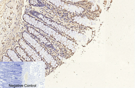
ARG66170 anti-beta Tubulin antibody IHC-P image
Immunohistochemistry: Paraffin-embedded Human colon tissue stained with ARG66170 anti-beta Tubulin antibody at 1:200 dilution (4°C, overnight). Antigen Retrieval: Boil tissue section in Sodium citrate buffer (pH 6.0) for 20 min.
Negative control was used by secondary antibody only.
-
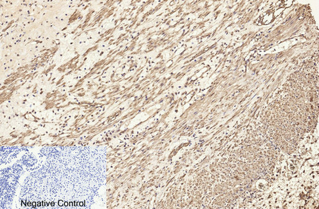
ARG66170 anti-beta Tubulin antibody IHC-P image
Immunohistochemistry: Paraffin-embedded Human Appendix tissue stained with ARG66170 anti-beta Tubulin antibody at 1:200 dilution (4°C, overnight). Antigen Retrieval: Boil tissue section in Sodium citrate buffer (pH 6.0) for 20 min.
Negative control was used by secondary antibody only.
-
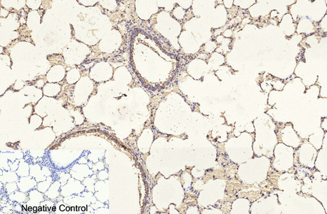
ARG66170 anti-beta Tubulin antibody IHC-P image
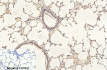
Immunohistochemistry: Paraffin-embedded Rat lung tissue stained with ARG66170 anti-beta Tubulin antibody at 1:200 dilution (4°C, overnight). Antigen Retrieval: Boil tissue section in Sodium citrate buffer (pH 6.0) for 20 min.
Negative control was used by secondary antibody only.
-

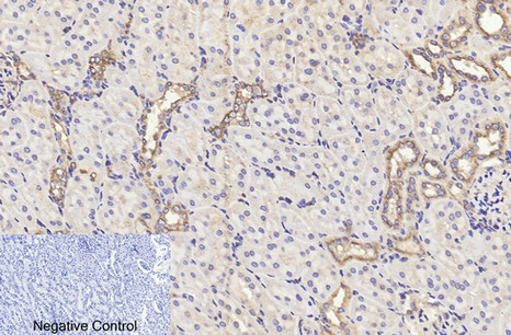
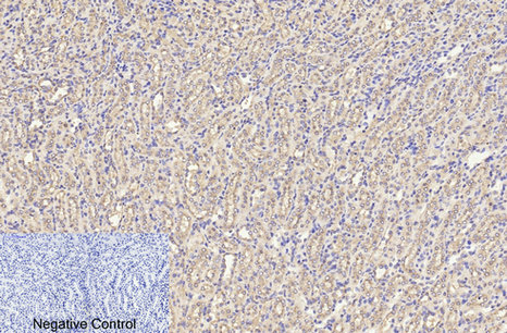
ARG66170 anti-beta Tubulin antibody IHC-P image
Immunohistochemistry: Paraffin-embedded Rat kidney tissue stained with ARG66170 anti-beta Tubulin antibody at 1:200 dilution (4°C, overnight). Antigen Retrieval: Boil tissue section in Sodium citrate buffer (pH 6.0) for 20 min.
Negative control was used by secondary antibody only.
-

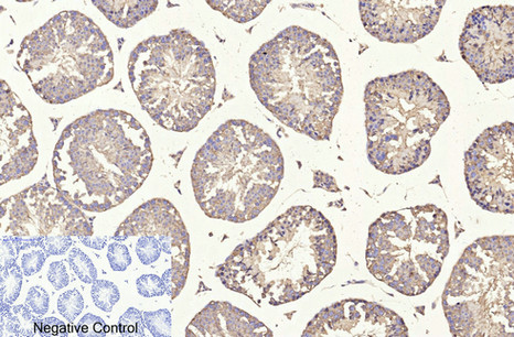
ARG66170 anti-beta Tubulin antibody IHC-P image
Immunohistochemistry: Paraffin-embedded Mouse testis tissue stained with ARG66170 anti-beta Tubulin antibody at 1:200 dilution (4°C, overnight). Antigen Retrieval: Boil tissue section in Sodium citrate buffer (pH 6.0) for 20 min.
Negative control was used by secondary antibody only.
-
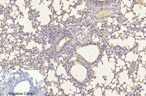
ARG66170 anti-beta Tubulin antibody IHC-P image
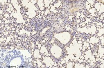
Immunohistochemistry: Paraffin-embedded Mouse lung tissue stained with ARG66170 anti-beta Tubulin antibody at 1:200 dilution (4°C, overnight). Antigen Retrieval: Boil tissue section in Sodium citrate buffer (pH 6.0) for 20 min.
Negative control was used by secondary antibody only.
-

ARG66170 anti-beta Tubulin antibody IHC-P image
Immunohistochemistry: Paraffin-embedded Mouse kidney tissue stained with ARG66170 anti-beta Tubulin antibody at 1:200 dilution (4°C, overnight). Antigen Retrieval: Boil tissue section in Sodium citrate buffer (pH 6.0) for 20 min.
Negative control was used by secondary antibody only.
-
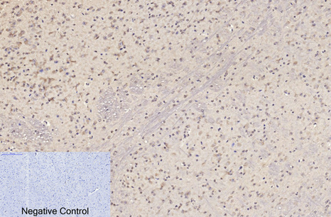
ARG66170 anti-beta Tubulin antibody IHC-P image
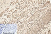
Immunohistochemistry: Paraffin-embedded Mouse brain tissue stained with ARG66170 anti-beta Tubulin antibody at 1:200 dilution (4°C, overnight). Antigen Retrieval: Boil tissue section in Sodium citrate buffer (pH 6.0) for 20 min.
Negative control was used by secondary antibody only.
-

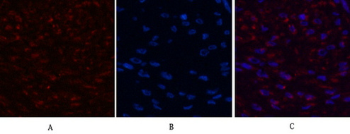
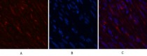
ARG66170 anti-beta Tubulin antibody IHC image
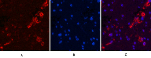
Immunohistochemistry: Human breast cancer tissue stained with ARG66170 anti-beta Tubulin antibody (red) at 1:200 dilution (4°C, overnight).
Picture A: Target. Picture B: DAPI. Picture C: merge of A+B.
-
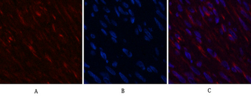
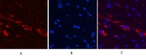
ARG66170 anti-beta Tubulin antibody IHC image
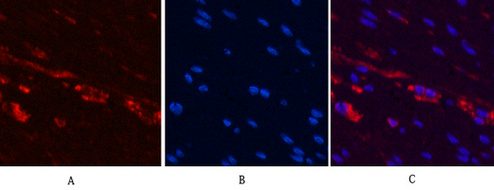
Immunohistochemistry: Human breast cancer tissue stained with ARG66170 anti-beta Tubulin antibody (red) at 1:200 dilution (4°C, overnight).
Picture A: Target. Picture B: DAPI. Picture C: merge of A+B.
-

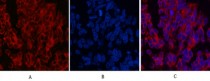
ARG66170 anti-beta Tubulin antibody IHC image
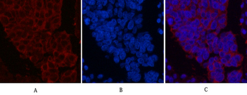
Immunohistochemistry: Human colon cancer tissue stained with ARG66170 anti-beta Tubulin antibody (red) at 1:200 dilution (4°C, overnight).
Picture A: Target. Picture B: DAPI. Picture C: merge of A+B.
-
ARG66170 anti-beta Tubulin antibody IHC image
Immunohistochemistry: Human colon cancer tissue stained with ARG66170 anti-beta Tubulin antibody (red) at 1:200 dilution (4°C, overnight).
Picture A: Target. Picture B: DAPI. Picture C: merge of A+B.
-
ARG66170 anti-beta Tubulin antibody IHC image
Immunohistochemistry: Human lung cancer tissue stained with ARG66170 anti-beta Tubulin antibody (red) at 1:200 dilution (4°C, overnight).
Picture A: Target. Picture B: DAPI. Picture C: merge of A+B.
-

ARG66170 anti-beta Tubulin antibody IHC image
Immunohistochemistry: Human lung cancer tissue stained with ARG66170 anti-beta Tubulin antibody (red) at 1:200 dilution (4°C, overnight).
Picture A: Target. Picture B: DAPI. Picture C: merge of A+B.