ARG52651
anti-beta Catenin antibody
anti-beta Catenin antibody for ICC/IF,IHC-Formalin-fixed paraffin-embedded sections,IHC-Frozen sections,Immunoprecipitation,Western blot and Human,Mouse,Rat
Cancer antibody; Cell Biology and Cellular Response antibody; Developmental Biology antibody; Neuroscience antibody; Signaling Transduction antibody

Overview
| Product Description | Rabbit Polyclonal antibody recognizes beta Catenin |
|---|---|
| Tested Reactivity | Hu, Ms, Rat |
| Tested Application | ICC/IF, IHC-Fr, IHC-P, IP, WB |
| Host | Rabbit |
| Clonality | Polyclonal |
| Isotype | IgG |
| Target Name | beta Catenin |
| Antigen Species | Human |
| Immunogen | Synthetic peptide from C-terminus (768-781) of the beta catenin protein. |
| Conjugation | Un-conjugated |
| Alternate Names | CTNNB; armadillo; MRD19; Catenin beta-1; Beta-catenin |
Application Instructions
| Application Suggestion |
|
||||||||||||
|---|---|---|---|---|---|---|---|---|---|---|---|---|---|
| Application Note | IHC-P: Antigen Retrieval: Boil tissue section in 10mM citrate buffer, pH 6.0 for 10 min followed by cooling at RT for 20 min. Incubation Time: 10 min at RT. * The dilutions indicate recommended starting dilutions and the optimal dilutions or concentrations should be determined by the scientist. |
||||||||||||
| Positive Control | Breast Carcinoma, A431 |
Properties
| Form | Liquid |
|---|---|
| Purification | Immunogen affinity purified |
| Buffer | PBS (pH 7.6), 1% BSA and < 0.1% Sodium azide |
| Preservative | < 0.1% Sodium azide |
| Stabilizer | 1% BSA |
| Storage Instruction | For continuous use, store undiluted antibody at 2-8°C for up to a week. For long-term storage, aliquot and store at -20°C or below. Storage in frost free freezers is not recommended. Avoid repeated freeze/thaw cycles. Suggest spin the vial prior to opening. The antibody solution should be gently mixed before use. |
| Note | For laboratory research only, not for drug, diagnostic or other use. |
Bioinformation
| Database Links | |
|---|---|
| Background | The catenins (α, β and γ) are ubiquitously expressed, cytoplasmic proteins associated with E-cadherin at cellular junctions. β-catenin also binds to N-cadherin and co-immunoprecipitates with APC. Cadherin/catenin complexes are linked to the cytoskeleton via a direct association between α-actinin and α-catenin. Increases tyrosine phosphorylation can disrupt catenin-cadherin complexes, influencing cellular adhesion. |
| Cellular Localization | Cytoplasm, Membrane |
| Highlight | Related Antibody Duos and Panels: ARG30144 Phospho beta Catenin Antibody Panel (Total, pS33, pS37, pT41/pS45) Related products: beta Catenin antibodies; beta Catenin Duos / Panels; Anti-Rabbit IgG secondary antibodies; Related news: Besides tumor suppression, what’s p53 busy for during embryogenesis? Wnt / beta-catenin signaling for gastric fundus specification |
| Research Area | Cancer antibody; Cell Biology and Cellular Response antibody; Developmental Biology antibody; Neuroscience antibody; Signaling Transduction antibody |
| Calculated MW | 85 kDa |
| PTM | Phosphorylation at Ser-552 by AMPK promotes stabilizion of the protein, enhancing TCF/LEF-mediated transcription (By similarity). Phosphorylation by GSK3B requires prior phosphorylation of Ser-45 by another kinase. Phosphorylation proceeds then from Thr-41 to Ser-37 and Ser-33. Phosphorylated by NEK2. EGF stimulates tyrosine phosphorylation. Phosphorylation on Tyr-654 decreases CDH1 binding and enhances TBP binding. Phosphorylated on Ser-33 and Ser-37 by HIPK2 and GSK3B, this phosphorylation triggers proteasomal degradation (PubMed:25169422). Phosphorylation on Ser-191 and Ser-246 by CDK5. Phosphorylation by CDK2 regulates insulin internalization. Phosphorylation by PTK6 at Tyr-64, Tyr-142, Tyr-331 and/or Tyr-333 with the predominant site at Tyr-64 is not essential for inhibition of transcriptional activity. Ubiquitinated by the SCF(BTRC) E3 ligase complex when phosphorylated by GSK3B, leading to its degradation. Ubiquitinated by a E3 ubiquitin ligase complex containing UBE2D1, SIAH1, CACYBP/SIP, SKP1, APC and TBL1X, leading to its subsequent proteasomal degradation (By similarity). S-nitrosylation at Cys-619 within adherens junctions promotes VEGF-induced, NO-dependent endothelial cell permeability by disrupting interaction with E-cadherin, thus mediating disassembly adherens junctions. O-glycosylation at Ser-23 decreases nuclear localization and transcriptional activity, and increases localization to the plasma membrane and interaction with E-cadherin CDH1. Deacetylated at Lys-49 by SIRT1. |
Images (2) Click the Picture to Zoom In
-
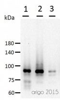
ARG52651 anti-beta Catenin antibody WB image
Western blot: 30 µg of 1) 293T, 2) 3T3, and 3) Mouse liver lysate stained with ARG52651 anti-beta Catenin antibody at 1:500 dilution.
-
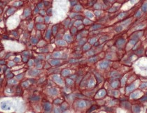
ARG52651 anti-Catenin-beta antibody IHC-P image
Immunohistochemistry: Human Breast Carcinoma stained with Catenin-beta antibody (ARG52651)


Customer's Feedback
Good

Review for anti-beta Catenin antibody
Application:WB
Sample:Rat liver
Sample Loading Amount:30 µg
Primary Antibody Dilution Factor:1:500
Primary Antibody Incubation Time:overnight
Primary Antibody Incubation Temperature:4 ºC

Specific References
Diuretic Action of Apelin-13 Mediated by Inhibiting cAMP/PKA/sPRR Pathway
WB / Mouse


Narasin inhibits tumor metastasis and growth of ERα‑positive breast cancer cells by inactivation of the TGF‑β/SMAD3 and IL‑6/STAT3 signaling pathways
WB / Human
Ubiquitin specific peptidase 5 enhances STAT3 signaling and promotes migration and invasion in Pancreatic Cancer.
WB / Human


