ARG40558
anti-beta Catenin antibody
anti-beta Catenin antibody for Flow cytometry,ICC/IF,IHC-Formalin-fixed paraffin-embedded sections,Western blot and Human,Mouse,Rat
Overview
| Product Description | Rabbit Polyclonal antibody recognizes beta Catenin |
|---|---|
| Tested Reactivity | Hu, Ms, Rat |
| Tested Application | FACS, ICC/IF, IHC-P, WB |
| Host | Rabbit |
| Clonality | Polyclonal |
| Isotype | IgG |
| Target Name | beta Catenin |
| Antigen Species | Human |
| Immunogen | Recombinant protein corresponding to A2-K233 of Human beta Catenin. |
| Conjugation | Un-conjugated |
| Alternate Names | CTNNB; armadillo; MRD19; Catenin beta-1; Beta-catenin |
Application Instructions
| Application Suggestion |
|
||||||||||
|---|---|---|---|---|---|---|---|---|---|---|---|
| Application Note | IHC-P: Antigen Retrieval: Heat mediation was performed in Citrate buffer (pH 6.0) for 20 min. * The dilutions indicate recommended starting dilutions and the optimal dilutions or concentrations should be determined by the scientist. |
Properties
| Form | Liquid |
|---|---|
| Purification | Affinity purification with immunogen. |
| Buffer | 0.2% Na2HPO4, 0.9% NaCl, 0.05% Sodium azide and 5% BSA. |
| Preservative | 0.05% Sodium azide |
| Stabilizer | 5% BSA |
| Concentration | 0.5 mg/ml |
| Storage Instruction | For continuous use, store undiluted antibody at 2-8°C for up to a week. For long-term storage, aliquot and store at -20°C or below. Storage in frost free freezers is not recommended. Avoid repeated freeze/thaw cycles. Suggest spin the vial prior to opening. The antibody solution should be gently mixed before use. |
| Note | For laboratory research only, not for drug, diagnostic or other use. |
Bioinformation
| Database Links | |
|---|---|
| Gene Symbol | CTNNB1 |
| Gene Full Name | catenin (cadherin-associated protein), beta 1, 88kDa |
| Background | The protein encoded by this gene is part of a complex of proteins that constitute adherens junctions (AJs). AJs are necessary for the creation and maintenance of epithelial cell layers by regulating cell growth and adhesion between cells. The encoded protein also anchors the actin cytoskeleton and may be responsible for transmitting the contact inhibition signal that causes cells to stop dividing once the epithelial sheet is complete. Finally, this protein binds to the product of the APC gene, which is mutated in adenomatous polyposis of the colon. Mutations in this gene are a cause of colorectal cancer (CRC), pilomatrixoma (PTR), medulloblastoma (MDB), and ovarian cancer. Three transcript variants encoding the same protein have been found for this gene.[provided by RefSeq, Oct 2009] |
| Function | Key downstream component of the canonical Wnt signaling pathway. In the absence of Wnt, forms a complex with AXIN1, AXIN2, APC, CSNK1A1 and GSK3B that promotes phosphorylation on N-terminal Ser and Thr residues and ubiquitination of CTNNB1 via BTRC and its subsequent degradation by the proteasome. In the presence of Wnt ligand, CTNNB1 is not ubiquitinated and accumulates in the nucleus, where it acts as a coactivator for transcription factors of the TCF/LEF family, leading to activate Wnt responsive genes. Involved in the regulation of cell adhesion. Acts as a negative regulator of centrosome cohesion. Involved in the CDK2/PTPN6/CTNNB1/CEACAM1 pathway of insulin internalization. Blocks anoikis of malignant kidney and intestinal epithelial cells and promotes their anchorage-independent growth by down-regulating DAPK2. Disrupts PML function and PML-NB formation by inhibiting RANBP2-mediated sumoylation of PML. Promotes neurogenesis by maintaining sympathetic neuroblasts within the cell cycle (By similarity). [UniProt] |
| Cellular Localization | Cytoplasm. Nucleus. Cytoplasm, cytoskeleton. Cell junction, adherens junction. Cell junction. Cell membrane. Cytoplasm, cytoskeleton, microtubule organizing center, centrosome. Cytoplasm, cytoskeleton, spindle pole. Cell junction, synapse. Cytoplasm, cytoskeleton, cilium basal body. Note=Colocalized with RAPGEF2 and TJP1 at cell-cell contacts (By similarity). [UniProt] |
| Calculated MW | 85 kDa |
| PTM | Phosphorylation at Ser-552 by AMPK promotes stabilizion of the protein, enhancing TCF/LEF-mediated transcription (By similarity). Phosphorylation by GSK3B requires prior phosphorylation of Ser-45 by another kinase. Phosphorylation proceeds then from Thr-41 to Ser-37 and Ser-33. Phosphorylated by NEK2. EGF stimulates tyrosine phosphorylation. Phosphorylation on Tyr-654 decreases CDH1 binding and enhances TBP binding. Phosphorylated on Ser-33 and Ser-37 by HIPK2 and GSK3B, this phosphorylation triggers proteasomal degradation (PubMed:25169422). Phosphorylation on Ser-191 and Ser-246 by CDK5. Phosphorylation by CDK2 regulates insulin internalization. Phosphorylation by PTK6 at Tyr-64, Tyr-142, Tyr-331 and/or Tyr-333 with the predominant site at Tyr-64 is not essential for inhibition of transcriptional activity. Ubiquitinated by the SCF(BTRC) E3 ligase complex when phosphorylated by GSK3B, leading to its degradation. Ubiquitinated by a E3 ubiquitin ligase complex containing UBE2D1, SIAH1, CACYBP/SIP, SKP1, APC and TBL1X, leading to its subsequent proteasomal degradation (By similarity). S-nitrosylation at Cys-619 within adherens junctions promotes VEGF-induced, NO-dependent endothelial cell permeability by disrupting interaction with E-cadherin, thus mediating disassembly adherens junctions. O-glycosylation at Ser-23 decreases nuclear localization and transcriptional activity, and increases localization to the plasma membrane and interaction with E-cadherin CDH1. Deacetylated at Lys-49 by SIRT1. [UniProt] |
Images (15) Click the Picture to Zoom In
-
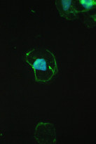
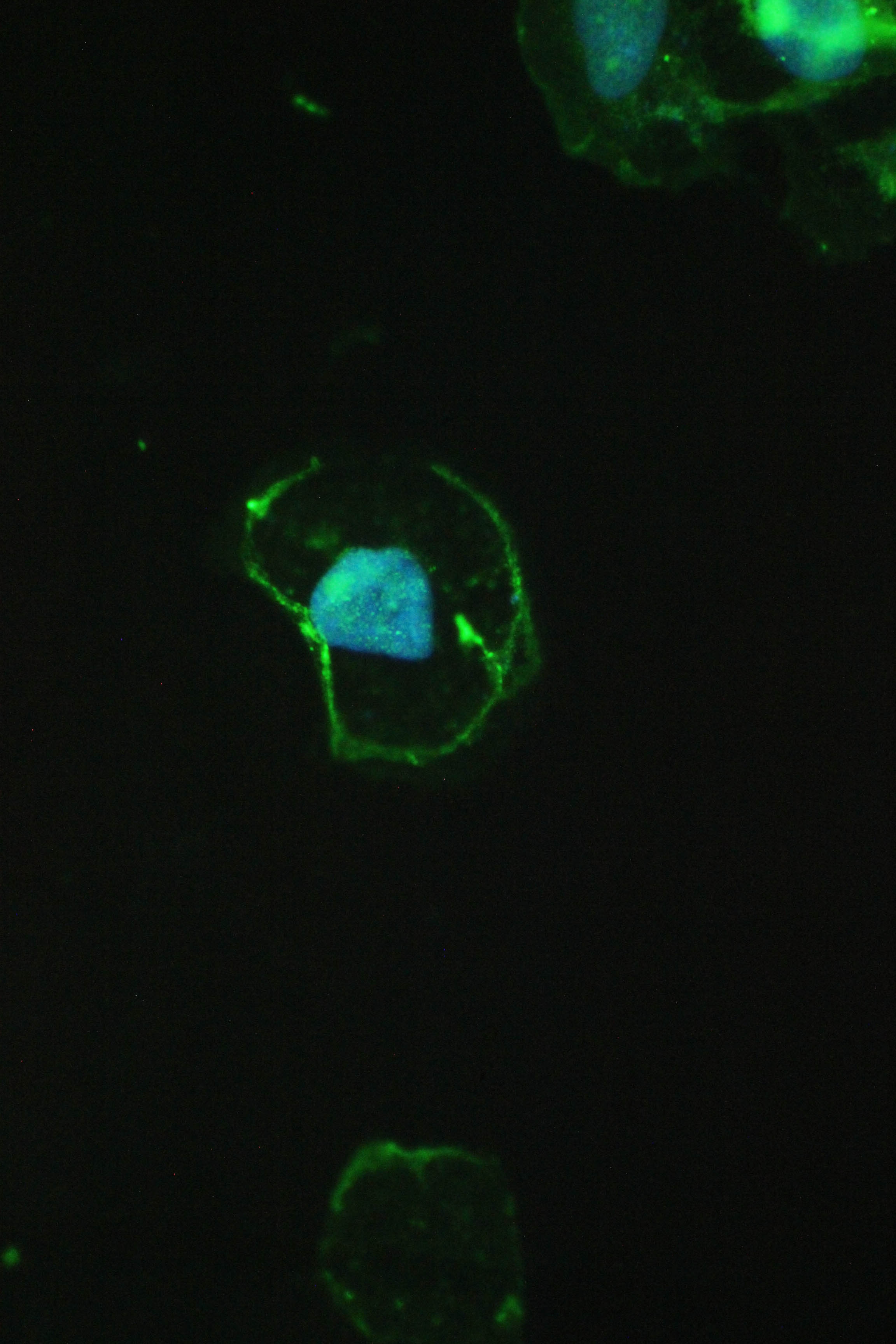
ARG40558 anti-beta Catenin antibody ICC/IF image
Immunofluorescence: A431 cells stained with ARG40558 anti-beta Catenin antibody (green) at 2 µg/ml, overnight at 4°C. DAPI (blue) for nuclear staining.
-
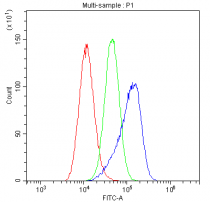
ARG40558 anti-beta Catenin antibody FACS image
Flow Cytometry: A549 cells were blocked with 10% normal goat serum and then stained with ARG40558 anti-beta Catenin antibody (blue) at 1 µg/10^6 cells for 30 min at 20°C, followed by incubation with DyLight®488 labelled secondary antibody. Isotype control antibody (green) was Rabbit IgG (1 µg/10^6 cells) used under the same conditions. Unlabelled sample (red) was also used as a control.
-
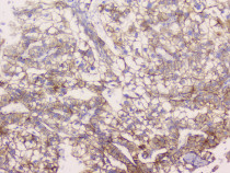
ARG40558 anti-beta Catenin antibody IHC-P image
Immunohistochemistry: Paraffin-embedded Human liver cancer tissue. Antigen Retrieval: Heat mediation was performed in Citrate buffer (pH 6.0, epitope retrieval solution) for 20 min. The tissue section was blocked with 10% goat serum. The tissue section was then stained with ARG40558 anti-beta Catenin antibody at 1 µg/ml, overnight at 4°C.
-
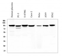
ARG40558 anti-beta Catenin antibody WB image
Western blot: 50 µg of samples under reducing conditions. Human placenta, PC-3, U-87MG, Caco-2, HeLa, A549 and K562 whole cell lysates stained with ARG40558 anti-beta Catenin antibody at 0.5 µg/ml, overnight at 4°C.
-
ARG40558 anti-beta Catenin antibody IHC-P image
Immunohistochemistry: Paraffin-embedded Human mammary cancer tissue. Antigen Retrieval: Heat mediation was performed in Citrate buffer (pH 6.0, epitope retrieval solution) for 20 min. The tissue section was blocked with 10% goat serum. The tissue section was then stained with ARG40558 anti-beta Catenin antibody at 1 µg/ml, overnight at 4°C.
-
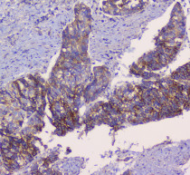
ARG40558 anti-beta Catenin antibody IHC-P image
Immunohistochemistry: Paraffin-embedded Human prostatic cancer tissue. Antigen Retrieval: Heat mediation was performed in Citrate buffer (pH 6.0, epitope retrieval solution) for 20 min. The tissue section was blocked with 10% goat serum. The tissue section was then stained with ARG40558 anti-beta Catenin antibody at 1 µg/ml, overnight at 4°C.
-
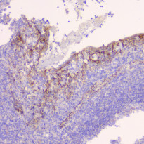
ARG40558 anti-beta Catenin antibody IHC-P image
Immunohistochemistry: Paraffin-embedded Human tonsil tissue. Antigen Retrieval: Heat mediation was performed in Citrate buffer (pH 6.0, epitope retrieval solution) for 20 min. The tissue section was blocked with 10% goat serum. The tissue section was then stained with ARG40558 anti-beta Catenin antibody at 1 µg/ml, overnight at 4°C.
-
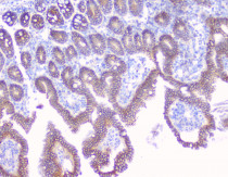
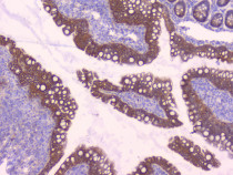
ARG40558 anti-beta Catenin antibody IHC-P image
Immunohistochemistry: Paraffin-embedded Mouse intestine tissue. Antigen Retrieval: Heat mediation was performed in Citrate buffer (pH 6.0, epitope retrieval solution) for 20 min. The tissue section was blocked with 10% goat serum. The tissue section was then stained with ARG40558 anti-beta Catenin antibody at 1 µg/ml, overnight at 4°C.
-
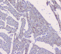
ARG40558 anti-beta Catenin antibody IHC-P image
Immunohistochemistry: Paraffin-embedded Mouse heart tissue. Antigen Retrieval: Heat mediation was performed in Citrate buffer (pH 6.0, epitope retrieval solution) for 20 min. The tissue section was blocked with 10% goat serum. The tissue section was then stained with ARG40558 anti-beta Catenin antibody at 1 µg/ml, overnight at 4°C.
-
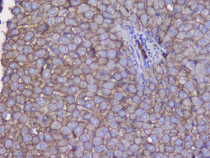
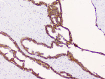
ARG40558 anti-beta Catenin antibody IHC-P image
Immunohistochemistry: Paraffin-embedded Mouse liver tissue. Antigen Retrieval: Heat mediation was performed in Citrate buffer (pH 6.0, epitope retrieval solution) for 20 min. The tissue section was blocked with 10% goat serum. The tissue section was then stained with ARG40558 anti-beta Catenin antibody at 1 µg/ml, overnight at 4°C.
-
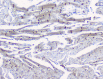
ARG40558 anti-beta Catenin antibody IHC-P image
Immunohistochemistry: Paraffin-embedded Rat heart tissue. Antigen Retrieval: Heat mediation was performed in Citrate buffer (pH 6.0, epitope retrieval solution) for 20 min. The tissue section was blocked with 10% goat serum. The tissue section was then stained with ARG40558 anti-beta Catenin antibody at 1 µg/ml, overnight at 4°C.
-
ARG40558 anti-beta Catenin antibody IHC-P image
Immunohistochemistry: Paraffin-embedded Rat intestine tissue. Antigen Retrieval: Heat mediation was performed in Citrate buffer (pH 6.0, epitope retrieval solution) for 20 min. The tissue section was blocked with 10% goat serum. The tissue section was then stained with ARG40558 anti-beta Catenin antibody at 1 µg/ml, overnight at 4°C.
-
ARG40558 anti-beta Catenin antibody IHC-P image
Immunohistochemistry: Paraffin-embedded Rat liver tissue. Antigen Retrieval: Heat mediation was performed in Citrate buffer (pH 6.0, epitope retrieval solution) for 20 min. The tissue section was blocked with 10% goat serum. The tissue section was then stained with ARG40558 anti-beta Catenin antibody at 1 µg/ml, overnight at 4°C.
-
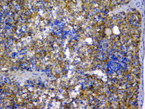
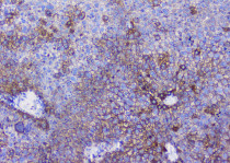
ARG40558 anti-beta Catenin antibody IHC-P image
Immunohistochemistry: Paraffin-embedded Rat spleen tissue. Antigen Retrieval: Heat mediation was performed in Citrate buffer (pH 6.0, epitope retrieval solution) for 20 min. The tissue section was blocked with 10% goat serum. The tissue section was then stained with ARG40558 anti-beta Catenin antibody at 1 µg/ml, overnight at 4°C.
-
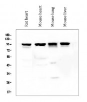
ARG40558 anti-beta Catenin antibody WB image
Western blot: 50 µg of samples under reducing conditions. Rat heart, Mouse heart, Mouse lung and Mouse liver lysates stained with ARG40558 anti-beta Catenin antibody at 0.5 µg/ml, overnight at 4°C.