ARG62670
anti-alpha Tubulin antibody [TU-01]
anti-alpha Tubulin antibody [TU-01] for ICC/IF,IHC-Formalin-fixed paraffin-embedded sections,Immunoprecipitation,Western blot and Human,Mouse,Arabidopsis,Eisenia,Nicotiana,Paramecium,Pig,Turkey,Yeast
Controls and Markers antibody; Neuroscience antibody; Signaling Transduction antibody
Overview
| Product Description | Mouse Monoclonal antibody [TU-01] recognizes alpha Tubulin |
|---|---|
| Tested Reactivity | Hu, Ms, Arabi, Eisenia, Nicotiana, Paramecium, Pig, Turkey, Yeast |
| Tested Application | ICC/IF, IHC-P, IP, WB |
| Specificity | The clone TU-01 recognizes the defined epitope (aa 65-97) on N-terminal structural domain of alpha-Tubulin. |
| Host | Mouse |
| Clonality | Monoclonal |
| Clone | TU-01 |
| Isotype | IgG1 |
| Target Name | alpha Tubulin |
| Antigen Species | Pig |
| Immunogen | Fraction of Tubulin purified from porcine brain by two cycles of polymerization - depolymerization. |
| Conjugation | Un-conjugated |
| Alternate Names | TUBA1; ALS22; Tubulin alpha-4A chain; Testis-specific alpha-tubulin; Alpha-tubulin 1; Tubulin alpha-1 chain; Tubulin H2-alpha; H2-ALPHA |
Application Instructions
| Application Suggestion |
|
||||||||||
|---|---|---|---|---|---|---|---|---|---|---|---|
| Application Note | WB: Sample preparation: Resuspend approx. 50 mil. cells in 1 ml cold Lysis buffer (1% laurylmaltoside in 20 mM Tris/Cl, 100 mM NaCl pH 8.2, 50 mM NaF including Protease inhibitor Cocktail). Incubate 60 min on ice. Centrifuge to remove cell debris. Mix lysate with reducing Laemmli SDS-PAGE sample buffer. ICC/IF: We recommend not only fixing but also permeabilizing the cells. * The dilutions indicate recommended starting dilutions and the optimal dilutions or concentrations should be determined by the scientist. |
||||||||||
| Positive Control | WB: HPB-ALL Human peripheral blood leukemia cell line (incubated for 60 min) and Porcine brain (incubated for 90 min). IHC-P: heart |
Properties
| Form | Liquid |
|---|---|
| Purification | Purified from ascites by precipitation methods. |
| Purity | > 95% (by SDS-PAGE) |
| Buffer | PBS (pH 7.4) and 15 mM Sodium azide |
| Preservative | 15 mM Sodium azide |
| Concentration | 1 mg/ml |
| Storage Instruction | For continuous use, store undiluted antibody at 2-8°C for up to a week. For long-term storage, aliquot and store at -20°C or below. Storage in frost free freezers is not recommended. Avoid repeated freeze/thaw cycles. Suggest spin the vial prior to opening. The antibody solution should be gently mixed before use. |
| Note | For laboratory research only, not for drug, diagnostic or other use. |
Bioinformation
| Database Links | |
|---|---|
| Background | The microtubules are intracellular dynamic polymers made up of evolutionarily conserved polymorphic alpha/beta-Tubulin heterodimers and a large number of microtubule-associated proteins (MAPs). The microtubules consist of 13 protofilaments and have an outer diameter 25 nm. Microtubules have their intrinsic polarity; highly dynamic plus ends and less dynamic minus ends. Microtubules are required for vital processes in eukaryotic cells including mitosis, meiosis, maintenance of cell shape and intracellular transport. Microtubules are also necessary for movement of cells by means of flagella and cilia. In mammalian tissue culture cells microtubules have their minus ends anchored in microtubule organizing centers (MTOCs).The GTP (guanosintriphosphate) molecule is an essential for Tubulin heterodimer to associate with other heterodimers to form microtubule. In vivo, microtubule dynamics vary considerably. Microtubule polymerization is reversible and a populations of microtubules in cells are on their minus ends either growing or shortening – this phenomenon is called dynamic instability of microtubules. On a practical level, microtubules can easily be stabilized by the addition of non-hydrolysable analogues of GTP (eg. GMPPCP) or more commonly by anti-cancer drugs such as Taxol. Taxol stabilizes microtubules at room temperature for many hours. Using limited proteolysis by enzymes both Tubulin subunits can be divided into N-terminal and C-terminal structural domains. The alpha-Tubulin (relative molecular weight around 50 kDa) is globular protein that exists in cells as part of soluble alpha/beta-Tubulin dimer or it is polymerized into microtubules. In different species it is coded by multiple Tubulin genes that form Tubulin classes (in human 6 genes). Expressed Tubulin genes are named Tubulin isotypes. Some of the Tubulin isotypes are expressed ubiquitously, while some have more restricted tissue expression. Alpha-Tubulin is also subject of numerous post-translational modifications. Tubulin isotypes and their posttranslational modifications are responsible for multiple Tubulin charge variants - Tubulin isoforms. Heterogeneity of alpha-Tubulin is concentrated in C-terminal structural domain._x000D_ |
| Research Area | Controls and Markers antibody; Neuroscience antibody; Signaling Transduction antibody |
| Calculated MW | 50 kDa |
| PTM | Some glutamate residues at the C-terminus are polyglutamylated, resulting in polyglutamate chains on the gamma-carboxyl group (PubMed:26875866). Polyglutamylation plays a key role in microtubule severing by spastin (SPAST). SPAST preferentially recognizes and acts on microtubules decorated with short polyglutamate tails: severing activity by SPAST increases as the number of glutamates per tubulin rises from one to eight, but decreases beyond this glutamylation threshold (PubMed:26875866). Some glutamate residues at the C-terminus are monoglycylated but not polyglycylated due to the absence of functional TTLL10 in human. Monoglycylation is mainly limited to tubulin incorporated into axonemes (cilia and flagella). Both polyglutamylation and monoglycylation can coexist on the same protein on adjacent residues, and lowering glycylation levels increases polyglutamylation, and reciprocally. The precise function of monoglycylation is still unclear (Probable). Acetylation of alpha chains at Lys-40 is located inside the microtubule lumen. This modification has been correlated with increased microtubule stability, intracellular transport and ciliary assembly. Methylation of alpha chains at Lys-40 is found in mitotic microtubules and is required for normal mitosis and cytokinesis contributing to genomic stability. |
Images (6) Click the Picture to Zoom In
-

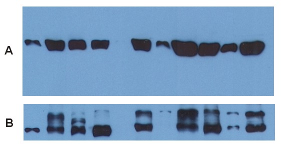
ARG62670 anti-alpha Tubulin antibody [TU-01] WB image
Western blot: Various cell lysates stained with ARG62670 anti-alpha Tubulin antibody [TU-01] as loading control.
-

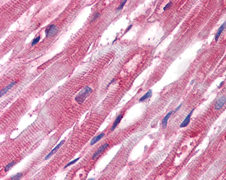
ARG62670 anti-alpha Tubulin antibody [TU-01] IHC-P image
Immunohistochemistry: Paraffin-embedded Human heart tissue stained with ARG62670 anti-alpha Tubulin antibody [TU-01].
-
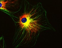
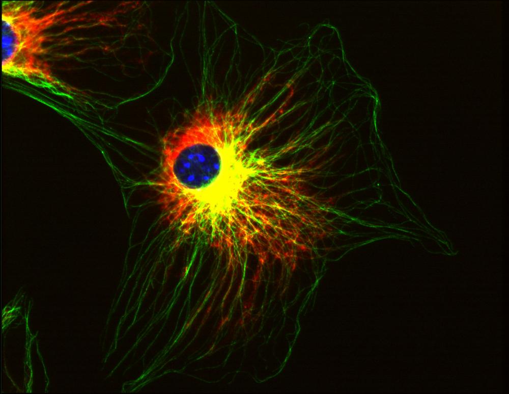
ARG62670 anti-alpha Tubulin antibody [TU-01] ICC/IF image
Immunofluorescence: 3T3 mouse embryonal fibroblast cells stained with ARG62670 anti-alpha Tubulin antibody [TU-01] (green).
Co-stained with Vimentin (red) and DAPI (blue). -
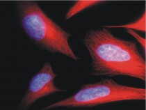
ARG62670 anti-alpha Tubulin antibody [TU-01] ICC/IF image
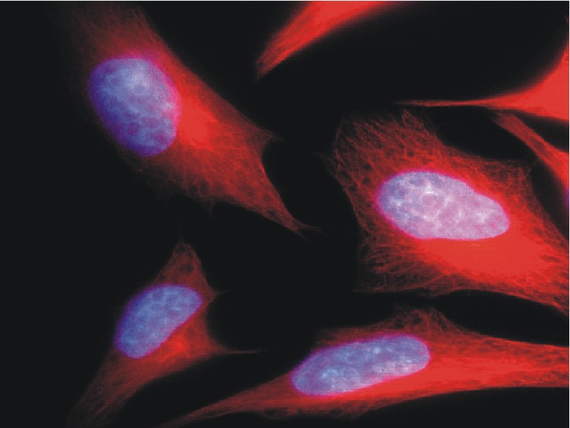
Immunofluorescence: HeLa cells stained with ARG62670 anti-alpha Tubulin antibody [TU-01] (red).
Cell nuclei was stained with DAPI (blue). -
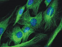
ARG62670 anti-alpha Tubulin antibody [TU-01] ICC/IF image
Immunofluorescence: 3T3 mouse embryonal fibroblast cells stained with ARG62670 anti-alpha Tubulin antibody [TU-01] (green).
Cell nuclei was stained with DAPI (blue). -

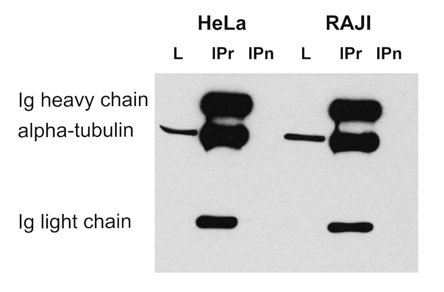
ARG62670 anti-alpha Tubulin antibody [TU-01] IP image
Immunoprecipitation: HeLa and Raji cell lysates immunoprecipitated by ARG62671 anti-alpha Tubulin antibody [TU-16] and stained with ARG62670 anti-alpha Tubulin antibody [TU-01]. IgM heavy chain (76-92 kDa) and IgM light chain (25-30 kDa) indicated. MW of alpha tubulin is around 50 kDa. L = lysate IPr = immunoprecipitate (reducing conditions) IPn = immunoprecipitate (non-reducing conditions).