ARG66788
anti-alpha Tubulin antibody
anti-alpha Tubulin antibody for ICC/IF,IHC-Formalin-fixed paraffin-embedded sections,Immunoprecipitation,Western blot and Human,Mouse,Rat
Overview
| Product Description | Mouse Monoclonal antibody recognizes alpha Tubulin |
|---|---|
| Tested Reactivity | Hu, Ms, Rat |
| Tested Application | ICC/IF, IHC-P, IP, WB |
| Host | Mouse |
| Clonality | Monoclonal |
| Isotype | IgG |
| Target Name | alpha Tubulin |
| Antigen Species | Human |
| Immunogen | Recombinant Protein of alpha Tubulin. |
| Conjugation | Un-conjugated |
| Alternate Names | CDCBM1; Tubulin beta-4 chain; Tubulin beta-3 chain; CFEOM3A; Tubulin beta-III; TUBB4; CDCBM; CFEOM3; FEOM3; beta-4 |
Application Instructions
| Application Suggestion |
|
||||||||||
|---|---|---|---|---|---|---|---|---|---|---|---|
| Application Note | IHC-P: Antigen Retrieval: Boil tissue section in Sodium citrate buffer (pH 6.0) for 20 min. * The dilutions indicate recommended starting dilutions and the optimal dilutions or concentrations should be determined by the scientist. |
||||||||||
| Positive Control | HeLa, Rat brian and Mouse brain | ||||||||||
| Observed Size | ~ 55 kDa |
Properties
| Form | Liquid |
|---|---|
| Purification | Affinity purification with immunogen. |
| Buffer | PBS (pH 7.4), 0.02% Sodium azide, 50% Glycerol and 0.5% BSA. |
| Preservative | 0.02% Sodium azide |
| Stabilizer | 50% Glycerol and 0.5% BSA |
| Storage Instruction | For continuous use, store undiluted antibody at 2-8°C for up to a week. For long-term storage, aliquot and store at -20°C. Storage in frost free freezers is not recommended. Avoid repeated freeze/thaw cycles. Suggest spin the vial prior to opening. The antibody solution should be gently mixed before use. |
| Note | For laboratory research only, not for drug, diagnostic or other use. |
Bioinformation
| Database Links | |
|---|---|
| Gene Symbol | TUBB3 |
| Gene Full Name | tubulin, beta 3 class III |
| Background | This gene encodes a class III member of the beta tubulin protein family. Beta tubulins are one of two core protein families (alpha and beta tubulins) that heterodimerize and assemble to form microtubules. This protein is primarily expressed in neurons and may be involved in neurogenesis and axon guidance and maintenance. Mutations in this gene are the cause of congenital fibrosis of the extraocular muscles type 3. Alternate splicing results in multiple transcript variants. A pseudogene of this gene is found on chromosome 6. [provided by RefSeq, Oct 2010] |
| Function | Tubulin is the major constituent of microtubules. It binds two moles of GTP, one at an exchangeable site on the beta chain and one at a non-exchangeable site on the alpha chain. TUBB3 plays a critical role in proper axon guidance and mantainance. Binding of NTN1/Netrin-1 to its receptor UNC5C might cause dissociation of UNC5C from polymerized TUBB3 in microtubules and thereby lead to increased microtubule dynamics and axon repulsion (PubMed:28483977). Plays a role in dorsal root ganglion axon projection towards the spinal cord (PubMed:28483977). [UniProt] |
| Cellular Localization | Cytoplasm, cytoskeleton. [UniProt] |
| Calculated MW | 50 kDa |
| PTM | Some glutamate residues at the C-terminus are polyglutamylated, resulting in polyglutamate chains on the gamma-carboxyl group (PubMed:26875866). Polyglutamylation plays a key role in microtubule severing by spastin (SPAST). SPAST preferentially recognizes and acts on microtubules decorated with short polyglutamate tails: severing activity by SPAST increases as the number of glutamates per tubulin rises from one to eight, but decreases beyond this glutamylation threshold (PubMed:26875866). Some glutamate residues at the C-terminus are monoglycylated but not polyglycylated due to the absence of functional TTLL10 in human. Monoglycylation is mainly limited to tubulin incorporated into axonemes (cilia and flagella). Both polyglutamylation and monoglycylation can coexist on the same protein on adjacent residues, and lowering glycylation levels increases polyglutamylation, and reciprocally. The precise function of monoglycylation is still unclear (Probable). Phosphorylated on Ser-172 by CDK1 during the cell cycle, from metaphase to telophase, but not in interphase. This phosphorylation inhibits tubulin incorporation into microtubules. [UniProt] |
Images (5) Click the Picture to Zoom In
-
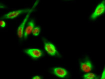
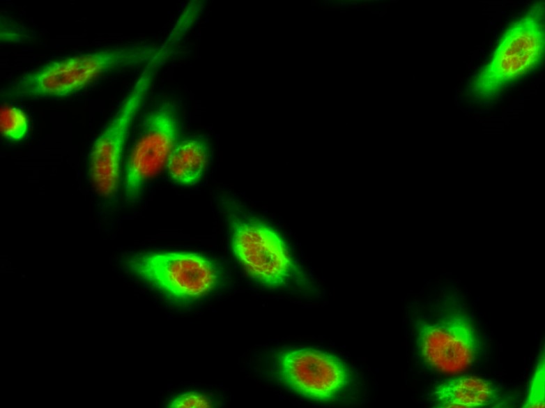
ARG66788 anti-alpha Tubulin antibody ICC/IF image
Immunofluorescence: HeLa cells stained with anti-DAPK3 phospho (Thr265) antibody (red) at 1:200 dilution, overnight at 4°C. Cells were co-stained with ARG66788 anti-alpha Tubulin antibody (green) at 1:200 dilution, overnight at 4°C.
-
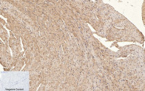
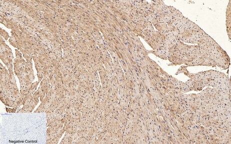
ARG66788 anti-alpha Tubulin antibody IHC-P image
Immunohistochemistry: Paraffin-embedded Mouse heart tissue. Antigen Retrieval: Boil tissue section in Sodium citrate buffer (pH 6.0) for 20 min. The tissue section was stained with ARG66788 anti-alpha Tubulin antibody at 1:200 dilution, overnight at 4°C. Negative control was used by secondary antibody only.
-
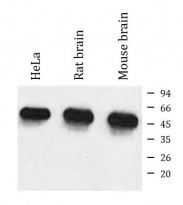
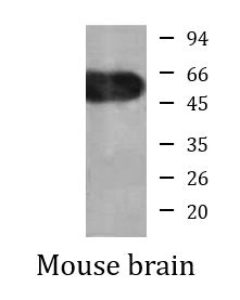
ARG66788 anti-alpha Tubulin antibody WB image
Western blot: HeLa, Rat brain and Mouse brain lysate stained with ARG66788 anti-alpha Tubulin antibody at 1:5000 dilution.
-

ARG66788 anti-alpha Tubulin antibody IP image
Immunoprecipitation: Mouse brain lysate were immunoprecipitated and stained with ARG66788 anti-alpha Tubulin antibody.
-
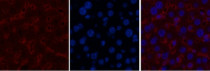
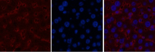
ARG66788 anti-alpha Tubulin antibody IHC-P image
Immunohistochemistry: Paraffin-embedded Mouse liver tissue stained with ARG66788 anti-alpha Tubulin antibody (red) at 1:200 dilution, overnight at 4°C. DAPI (blue) for nuclear staining.