ARG43207
anti-alpha Synuclein antibody [2A7]
anti-alpha Synuclein antibody [2A7] for ICC/IF,IHC-Frozen sections,IHC-Formalin-fixed paraffin-embedded sections,Western blot and Human,Mouse,Rat,Cow,Pig
Overview
| Product Description | Mouse Monoclonal antibody [2A7] recognizes alpha Synuclein |
|---|---|
| Tested Reactivity | Hu, Ms, Rat, Cow, Pig |
| Tested Application | ICC/IF, IHC-Fr, IHC-P, WB |
| Host | Mouse |
| Clonality | Monoclonal |
| Clone | 2A7 |
| Isotype | IgG1 |
| Target Name | alpha Synuclein |
| Antigen Species | Human |
| Immunogen | Recombinant full length Human alpha synuclein protein. |
| Conjugation | Un-conjugated |
| Alternate Names | Non-A4 component of amyloid precursor; Alpha-synuclein; PARK4; PARK1; PD1; NACP; Non-A beta component of AD amyloid |
Application Instructions
| Application Suggestion |
|
||||||||||
|---|---|---|---|---|---|---|---|---|---|---|---|
| Application Note | * The dilutions indicate recommended starting dilutions and the optimal dilutions or concentrations should be determined by the scientist. | ||||||||||
| Positive Control | Rat brain and Rat spinal cord | ||||||||||
| Observed Size | ~ 17 kDa |
Properties
| Form | Liquid |
|---|---|
| Purification | Purified |
| Buffer | PBS, 5 mM Sodium azide and 50% Glycerol. |
| Preservative | 5 mM Sodium azide |
| Stabilizer | 50% Glycerol |
| Concentration | 1 mg/ml |
| Storage Instruction | For continuous use, store undiluted antibody at 2-8°C for up to a week. For long-term storage, aliquot and store at -20°C. Storage in frost free freezers is not recommended. Avoid repeated freeze/thaw cycles. Suggest spin the vial prior to opening. The antibody solution should be gently mixed before use. |
| Note | For laboratory research only, not for drug, diagnostic or other use. |
Bioinformation
| Database Links | |
|---|---|
| Gene Symbol | SNCA |
| Gene Full Name | synuclein, alpha (non A4 component of amyloid precursor) |
| Background | Alpha-synuclein is a member of the synuclein family, which also includes beta- and gamma-synuclein. Synucleins are abundantly expressed in the brain and alpha- and beta-synuclein inhibit phospholipase D2 selectively. SNCA may serve to integrate presynaptic signaling and membrane trafficking. Defects in SNCA have been implicated in the pathogenesis of Parkinson disease. SNCA peptides are a major component of amyloid plaques in the brains of patients with Alzheimer's disease. Alternatively spliced transcripts encoding different isoforms have been identified for this gene. [provided by RefSeq, Feb 2016] |
| Function | Neuronal protein that plays several roles in synaptic activity such as regulation of synaptic vesicle trafficking and subsequent neurotransmitter release. Participates as a monomer in synaptic vesicle exocytosis by enhancing vesicle priming, fusion and dilation of exocytotic fusion pores (PubMed:28288128, PubMed:30404828). Mechanistically, acts by increasing local Ca(2+) release from microdomains which is essential for the enhancement of ATP-induced exocytosis (PubMed:30404828). Acts also as a molecular chaperone in its multimeric membrane-bound state, assisting in the folding of synaptic fusion components called SNAREs (Soluble NSF Attachment Protein REceptors) at presynaptic plasma membrane in conjunction with cysteine string protein-alpha/DNAJC5 (PubMed:20798282). This chaperone activity is important to sustain normal SNARE-complex assembly during aging (PubMed:20798282). Plays also a role in the regulation of the dopamine neurotransmission by associating with the dopamine transporter (DAT1) and thereby modulating its activity (PubMed:26442590). [UniProt] |
| Cellular Localization | Cytoplasm, cytosol. Membrane. Nucleus. Cell junction, synapse. Secreted. Note=Membrane-bound in dopaminergic neurons. [UniProt] |
| Calculated MW | 14 kDa |
| PTM | Phosphorylated, predominantly on serine residues. Phosphorylation by CK1 appears to occur on residues distinct from the residue phosphorylated by other kinases. Phosphorylation of Ser-129 is selective and extensive in synucleinopathy lesions. In vitro, phosphorylation at Ser-129 promoted insoluble fibril formation. Phosphorylated on Tyr-125 by a PTK2B-dependent pathway upon osmotic stress. Hallmark lesions of neurodegenerative synucleinopathies contain alpha-synuclein that is modified by nitration of tyrosine residues and possibly by dityrosine cross-linking to generated stable oligomers. Ubiquitinated. The predominant conjugate is the diubiquitinated form (By similarity). Acetylation at Met-1 seems to be important for proper folding and native oligomeric structure. [UniProt] |
Images (5) Click the Picture to Zoom In
-

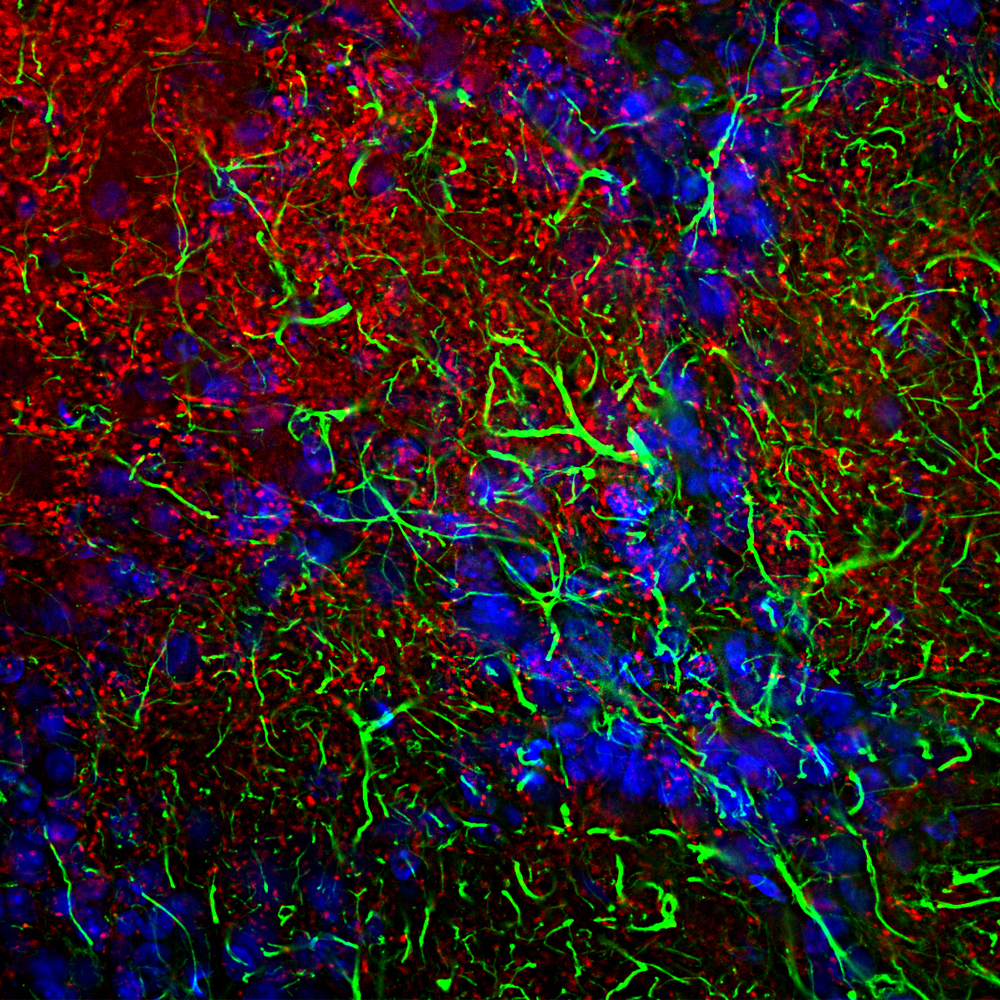
ARG43207 anti-alpha Synuclein antibody [2A7] IHC-Fr image
Immunohistochemistry: Frozen section of Rat cerebellum tissue stained with ARG43207 anti-alpha Synuclein antibody [2A7] (red) at 1:1000 dilution, and costained with anti-GFAP antibody (green) at 1:5000 dilution. Hoechst (blue) for nuclear staining. Sample preparation: Following transcardial perfusion of Rat with 4% paraformaldehyde, brain was post fixed for 24 hours, cut to 45 µM, and free-floating sections were stained with above antibodies.
The alpha synuclein protein is concentrated in synaptic regions, while the GFAP antibody stains the filamentous cytoskeleton of Bergmann glia and astrocytic cells.
-
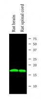
ARG43207 anti-alpha Synuclein antibody [2A7] WB image
Western blot: Rat brain and Rat spinal cord lysates stained with ARG43207 anti-alpha Synuclein antibody [2A7] (green) at 1:1000 dilution.
-

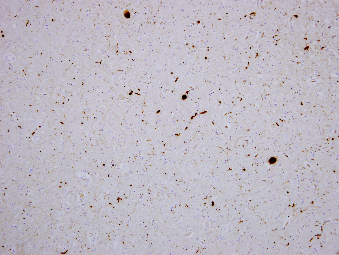
ARG43207 anti-alpha Synuclein antibody [2A7] IHC-P image
Immunohistochemistry: Paraffin-embedded cortex tissue of a patient with Parkinson's disease. Tissue section was stained with ARG43207 anti-alpha Synuclein antibody [2A7] at 1:1000 dilution. Antibody revealed with HRP and DAB.
The Lewy bodies and other typical inclusions of Parkinson's disease are seen in brown.
-
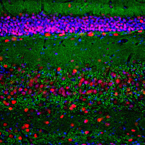
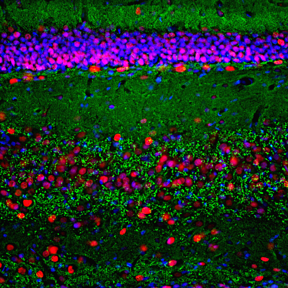
ARG43207 anti-alpha Synuclein antibody [2A7] IHC-Fr image
Immunohistochemistry: Frozen section of Rat hippocampus tissue stained with ARG43207 anti-alpha Synuclein antibody [2A7] (green) at 1:1000 dilution, and costained with anti-MeCP2 antibody (red) at 1:2000 dilution. DAPI (blue) for nuclear staining. Sample preparation: Following transcardial perfusion of Rat with 4% paraformaldehyde, brain was post fixed for 1 hour, cut to 45 µM, and free-floating sections were stained with above antibodies.
The alpha synuclein protein is concentrated in synaptic regions, and the MeCP2 antibody stains the nuclei of neuronal cells.
-

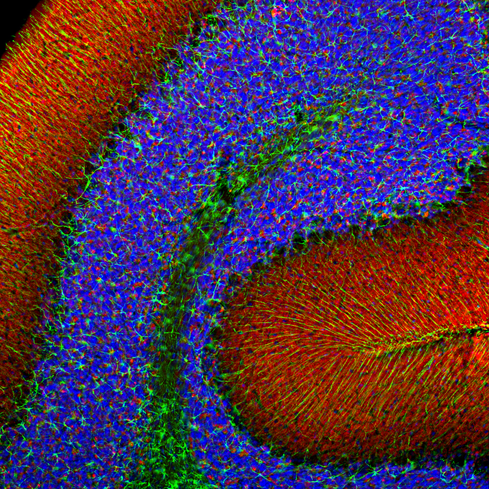
ARG43207 anti-alpha Synuclein antibody [2A7] IHC-Fr image
Immunohistochemistry: Frozen section of Rat olfactory bulb tissue stained with ARG43207 anti-alpha Synuclein antibody [2A7] (red) at 1:1000 dilution, and costained with anti-GFAP antibody (green) at 1:5000 dilution. DAPI (blue) for nuclear staining. Sample preparation: Following transcardial perfusion of Rat with 4% paraformaldehyde, brain was post fixed for 24 hours, cut to 45 µM, and free-floating sections were stained with above antibodies.
The alpha synuclein protein is concentrated in synaptic regions, while the GFAP antibody stains the filamentous backbone of astroglial cells.