ARG63953
anti-alpha Catenin antibody
anti-alpha Catenin antibody for IHC-Formalin-fixed paraffin-embedded sections,Western blot and Human
Cancer antibody; Developmental Biology antibody; Signaling Transduction antibody
Overview
| Product Description | Goat Polyclonal antibody recognizes alpha Catenin |
|---|---|
| Tested Reactivity | Hu |
| Predict Reactivity | Ms, Rat, Dog |
| Tested Application | IHC-P, WB |
| Host | Goat |
| Clonality | Polyclonal |
| Isotype | IgG |
| Target Name | alpha Catenin |
| Antigen Species | Human |
| Immunogen | C-KREKQDETQTKIK |
| Conjugation | Un-conjugated |
| Alternate Names | Catenin alpha-1; Renal carcinoma antigen NY-REN-13; Cadherin-associated protein; CAP102; Alpha E-catenin |
Application Instructions
| Application Suggestion |
|
||||||
|---|---|---|---|---|---|---|---|
| Application Note | WB: Recommend incubate at RT for 1h. IHC-P: Antigen Retrieval: Steam tissue section in Citrate buffer (pH 6.0). * The dilutions indicate recommended starting dilutions and the optimal dilutions or concentrations should be determined by the scientist. |
Properties
| Form | Liquid |
|---|---|
| Purification | Purified from goat serum by antigen affinity chromatography. |
| Buffer | Tris saline (pH 7.3), 0.02% Sodium azide and 0.5% BSA. |
| Preservative | 0.02% Sodium azide |
| Stabilizer | 0.5% BSA |
| Concentration | 0.5 mg/ml |
| Storage Instruction | For continuous use, store undiluted antibody at 2-8°C for up to a week. For long-term storage, aliquot and store at -20°C or below. Storage in frost free freezers is not recommended. Avoid repeated freeze/thaw cycles. Suggest spin the vial prior to opening. The antibody solution should be gently mixed before use. |
| Note | For laboratory research only, not for drug, diagnostic or other use. |
Bioinformation
| Database Links | |
|---|---|
| Gene Symbol | CTNNA1 |
| Gene Full Name | catenin (cadherin-associated protein), alpha 1, 102kDa |
| Function | Associates with the cytoplasmic domain of a variety of cadherins. The association of catenins to cadherins produces a complex which is linked to the actin filament network, and which seems to be of primary importance for cadherins cell-adhesion properties. Can associate with both E- and N-cadherins. Originally believed to be a stable component of E-cadherin/catenin adhesion complexes and to mediate the linkage of cadherins to the actin cytoskeleton at adherens junctions. In contrast, cortical actin was found to be much more dynamic than E-cadherin/catenin complexes and CTNNA1 was shown not to bind to F-actin when assembled in the complex suggesting a different linkage between actin and adherens junctions components. The homodimeric form may regulate actin filament assembly and inhibit actin branching by competing with the Arp2/3 complex for binding to actin filaments. May play a crucial role in cell differentiation. [UniProt] |
| Research Area | Cancer antibody; Developmental Biology antibody; Signaling Transduction antibody |
| Calculated MW | 100 kDa |
| PTM | Sumoylated. Phosphorylation seems to contribute to the strength of cell-cell adhesion rather than to the basic capacity for cell-cell adhesion. |
Images (4) Click the Picture to Zoom In
-
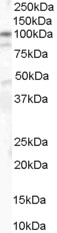
ARG63953 anti-alpha Catenin antibody WB image
Western blot: 35 µg of Human colon lysate stained with ARG63953 anti-alpha Catenin antibody at 1 µg/ml dilution.
-
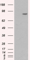
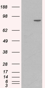
ARG63953 anti-alpha Catenin antibody WB image
Western blot: 1) Mock transfection and 2) Human CTNNA1 expressing plasmid transfected HEK293 cell lysate stained with ARG63953 anti-alpha Catenin antibody.
-
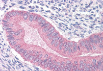
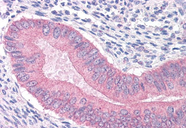
ARG63953 anti-alpha Catenin antibody IHC-P image
Immunohistochemistry: Paraffin-embedded Human uterus tissue. Antigen Retrieval: Steam tissue section in Citrate buffer (pH 6.0). The tissue section was stained with ARG63953 anti-alpha Catenin antibody at 3.75 µg/ml dilution followed by AP-staining.
-
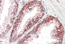
ARG63953 anti-alpha Catenin antibody IHC-P image
Immunohistochemistry: Paraffin-embedded Human Prostate (Steamed antigen retrieval with citrate buffer pH 6) stained with ARG63953 anti-alpha Catenin antibody at 3.8 µg/ml dilution, followed by AP-staining.