ARG58521
anti-alpha 1 Catenin antibody
anti-alpha 1 Catenin antibody for Flow cytometry,ICC/IF,IHC-Formalin-fixed paraffin-embedded sections,Western blot and Human,Mouse,Rat
Overview
| Product Description | Rabbit Polyclonal antibody recognizes alpha 1 Catenin |
|---|---|
| Tested Reactivity | Hu, Ms, Rat |
| Tested Application | FACS, ICC/IF, IHC-P, WB |
| Host | Rabbit |
| Clonality | Polyclonal |
| Isotype | IgG |
| Target Name | alpha 1 Catenin |
| Antigen Species | Human |
| Immunogen | Human CTNNA1 recombinant protein (Position: D143-D292). Human CTNNA1 shares 98% amino acid (aa) sequence identity with Mouse CTNNA1. |
| Conjugation | Un-conjugated |
| Alternate Names | Catenin alpha-1; Renal carcinoma antigen NY-REN-13; Cadherin-associated protein; CAP102; Alpha E-catenin |
Application Instructions
| Application Suggestion |
|
||||||||||
|---|---|---|---|---|---|---|---|---|---|---|---|
| Application Note | IHC-P: Antigen Retrieval: Heat mediation was performed in Citrate buffer (pH 6.0) for 20 min. * The dilutions indicate recommended starting dilutions and the optimal dilutions or concentrations should be determined by the scientist. |
Properties
| Form | Liquid |
|---|---|
| Purification | Affinity purification with immunogen. |
| Buffer | 0.9% NaCl, 0.2% Na2HPO4, 0.05% Sodium azide and 5% BSA. |
| Preservative | 0.05% Sodium azide |
| Stabilizer | 5% BSA |
| Concentration | 0.5 mg/ml |
| Storage Instruction | For continuous use, store undiluted antibody at 2-8°C for up to a week. For long-term storage, aliquot and store at -20°C or below. Storage in frost free freezers is not recommended. Avoid repeated freeze/thaw cycles. Suggest spin the vial prior to opening. The antibody solution should be gently mixed before use. |
| Note | For laboratory research only, not for drug, diagnostic or other use. |
Bioinformation
| Database Links | |
|---|---|
| Gene Symbol | CTNNA1 |
| Gene Full Name | catenin (cadherin-associated protein), alpha 1, 102kDa |
| Function | Associates with the cytoplasmic domain of a variety of cadherins. The association of catenins to cadherins produces a complex which is linked to the actin filament network, and which seems to be of primary importance for cadherins cell-adhesion properties. Can associate with both E- and N-cadherins. Originally believed to be a stable component of E-cadherin/catenin adhesion complexes and to mediate the linkage of cadherins to the actin cytoskeleton at adherens junctions. In contrast, cortical actin was found to be much more dynamic than E-cadherin/catenin complexes and CTNNA1 was shown not to bind to F-actin when assembled in the complex suggesting a different linkage between actin and adherens junctions components. The homodimeric form may regulate actin filament assembly and inhibit actin branching by competing with the Arp2/3 complex for binding to actin filaments. May play a crucial role in cell differentiation. [UniProt] |
| Cellular Localization | Isoform 1: Cytoplasm, cytoskeleton. Cell junction, adherens junction. Cell membrane; Peripheral membrane protein; Cytoplasmic side. Cell junction. Found at cell-cell boundaries and probably at cell-matrix boundaries. [UniProt] |
| Calculated MW | 100 kDa |
| PTM | Sumoylated. Phosphorylation seems to contribute to the strength of cell-cell adhesion rather than to the basic capacity for cell-cell adhesion. [UniProt] |
Images (7) Click the Picture to Zoom In
-

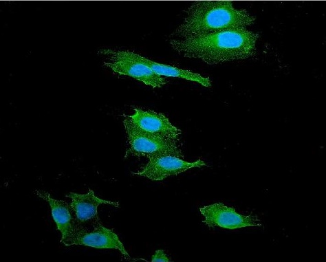
ARG58521 anti-alpha 1 Catenin antibody ICC/IF image
Immunofluorescence: U2OS cells were blocked with 10% goat serum and then stained with ARG58521 anti-alpha 1 Catenin antibody (green) at 2 µg/ml dilution, overnight at 4°C. DAPI (blue) for nuclear staining.
-
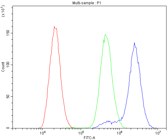
ARG58521 anti-alpha 1 Catenin antibody FACS image
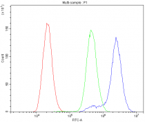
Flow Cytometry: U-87 cells were blocked with 10% normal goat serum, and then stained with ARG58521 anti-alpha 1 Catenin antibody (blue) at 1 µg/10^6 cells for 30 min at 20°C, followed by DyLight®488 labelled secondary antibody. Isotype control antibody (green) was rabbit IgG (1 µg/10^6 cells) used under the same conditions. Unlabelled sample (red) was also used as a control.
-
ARG58521 anti-alpha 1 Catenin antibody IHC-P image
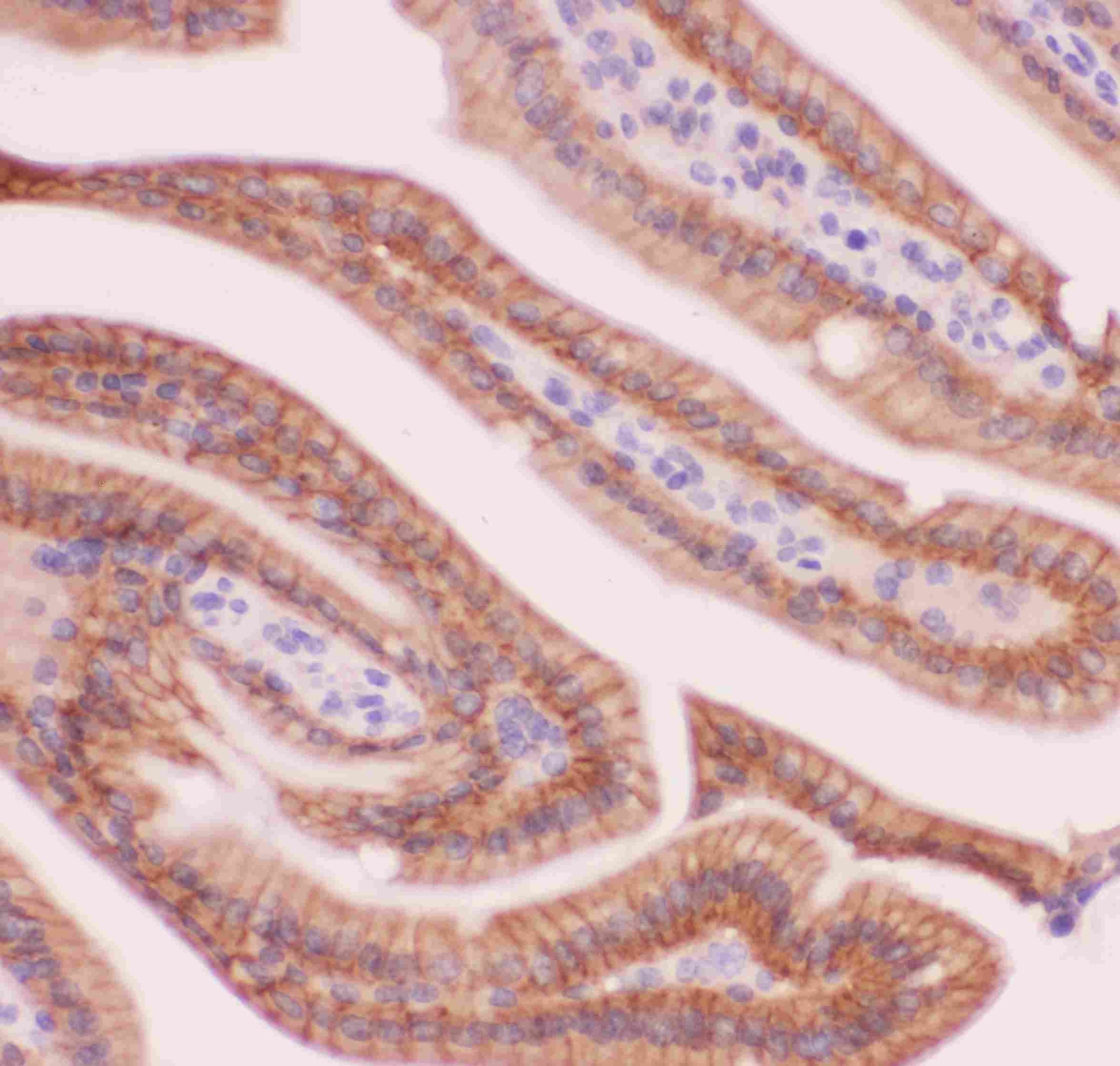
Immunohistochemistry: Paraffin-embedded Rat intestine tissue. Antigen Retrieval: Heat mediated was performed in Citrate buffer (pH 6.0, epitope retrieval solution) for 20 min. The tissue section was blocked with 10% goat serum. The tissue section was then stained with ARG58521 anti-alpha 1 Catenin antibody at 1 µg/ml dilution, overnight at 4°C.
-
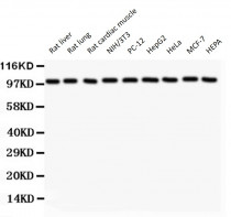
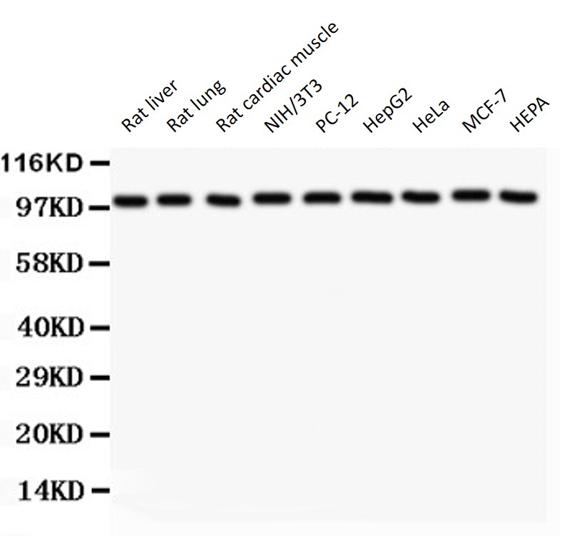
ARG58521 anti-alpha 1 Catenin antibody WB image
Western blot: 50 µg of samples under reducing conditions. Rat liver, Rat lung, Rat cardiac muscle, NIH/3T3, PC-12, HepG2, HeLa, MCF-7 and HEPA whole cell lysates stained with ARG58521 anti-alpha 1 Catenin antibody at 0.5 µg/ml, overnight at 4°C.
-
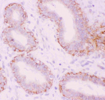
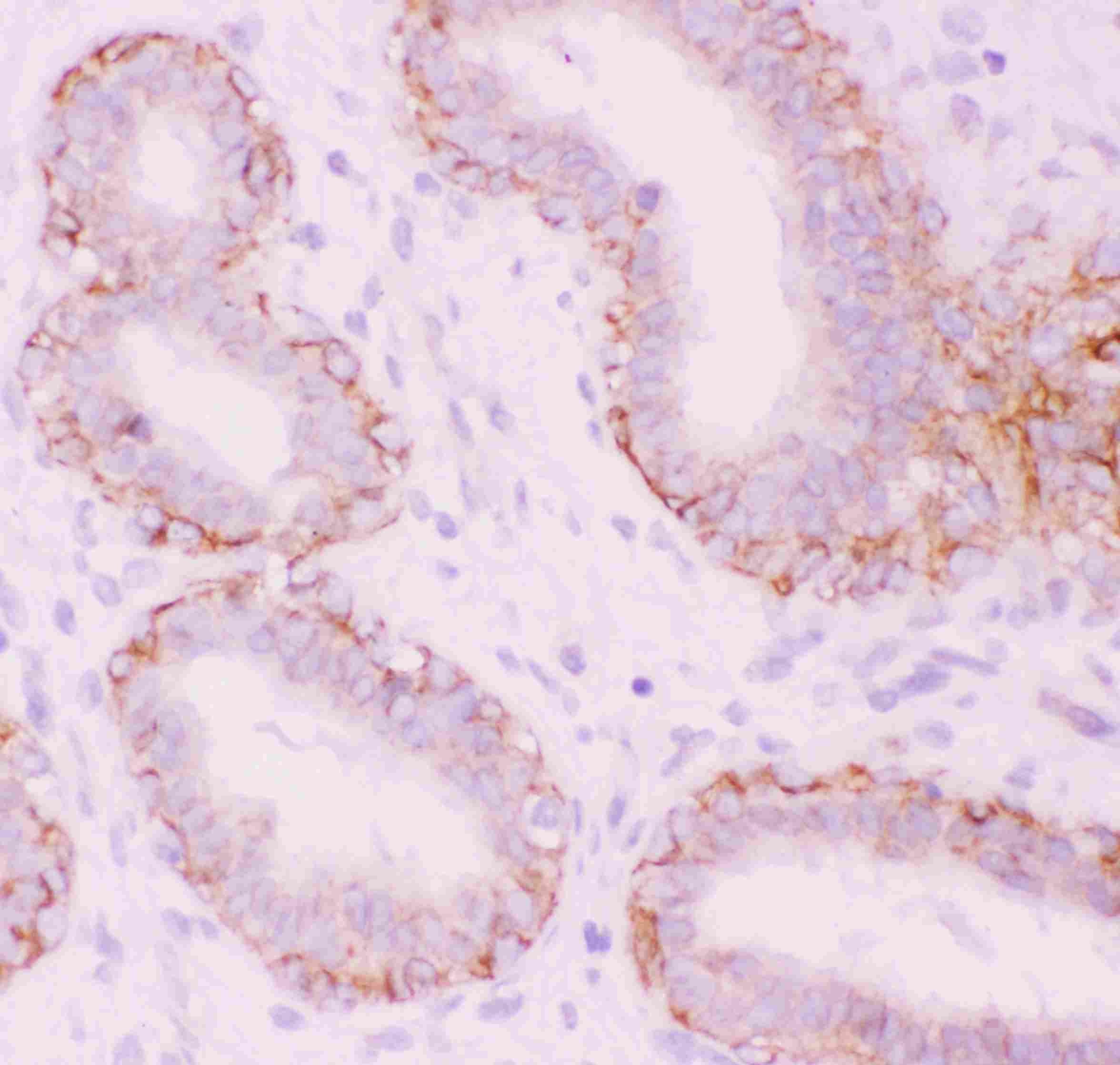
ARG58521 anti-alpha 1 Catenin antibody IHC-P image
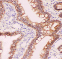
Immunohistochemistry: Paraffin-embedded Human mammary tissue. Antigen Retrieval: Heat mediated was performed in Citrate buffer (pH 6.0, epitope retrieval solution) for 20 min. The tissue section was blocked with 10% goat serum. The tissue section was then stained with ARG58521 anti-alpha 1 Catenin antibody at 1 µg/ml dilution, overnight at 4°C.
-
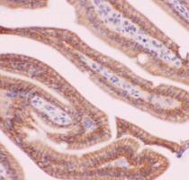
ARG58521 anti-alpha 1 Catenin antibody IHC-P image
Immunohistochemistry: Paraffin-embedded Mouse intestine tissue. Antigen Retrieval: Heat mediated was performed in Citrate buffer (pH 6.0, epitope retrieval solution) for 20 min. The tissue section was blocked with 10% goat serum. The tissue section was then stained with ARG58521 anti-alpha 1 Catenin antibody at 1 µg/ml dilution, overnight at 4°C.
-

ARG58521 anti-alpha 1 Catenin antibody WB image
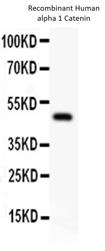
Western blot: 0.5 ng of Recombinant Human alpha 1 Catenin Protein stained with ARG58521 anti-alpha 1 Catenin antibody at 0.5 µg/ml, overnight at 4°C.