ARG55245
anti-WEE1 antibody
anti-WEE1 antibody for IHC-Formalin-fixed paraffin-embedded sections,Western blot and Human,Mouse,Rat
Cell Biology and Cellular Response antibody; Gene Regulation antibody
Overview
| Product Description | Rabbit Polyclonal antibody recognizes WEE1 |
|---|---|
| Tested Reactivity | Hu, Ms, Rat |
| Tested Application | IHC-P, WB |
| Host | Rabbit |
| Clonality | Polyclonal |
| Isotype | IgG |
| Target Name | WEE1 |
| Antigen Species | Human |
| Immunogen | KLH-conjugated synthetic peptide corresponding to aa. 144-173 (Center) of Human WEE1. |
| Conjugation | Un-conjugated |
| Alternate Names | Wee1A kinase; Wee1-like protein kinase; WEE1hu; WEE1A; EC 2.7.10.2 |
Application Instructions
| Application Suggestion |
|
||||||
|---|---|---|---|---|---|---|---|
| Application Note | * The dilutions indicate recommended starting dilutions and the optimal dilutions or concentrations should be determined by the scientist. |
Properties
| Form | Liquid |
|---|---|
| Purification | Purification with Protein G. |
| Buffer | PBS and 0.09% (W/V) Sodium azide |
| Preservative | 0.09% (W/V) Sodium azide |
| Storage Instruction | For continuous use, store undiluted antibody at 2-8°C for up to a week. For long-term storage, aliquot and store at -20°C or below. Storage in frost free freezers is not recommended. Avoid repeated freeze/thaw cycles. Suggest spin the vial prior to opening. The antibody solution should be gently mixed before use. |
| Note | For laboratory research only, not for drug, diagnostic or other use. |
Bioinformation
| Database Links | |
|---|---|
| Gene Symbol | WEE1 |
| Gene Full Name | WEE1 G2 checkpoint kinase |
| Background | This gene encodes a nuclear protein, which is a tyrosine kinase belonging to the Ser/Thr family of protein kinases. This protein catalyzes the inhibitory tyrosine phosphorylation of CDC2/cyclin B kinase, and appears to coordinate the transition between DNA replication and mitosis by protecting the nucleus from cytoplasmically activated CDC2 kinase. [provided by RefSeq, Jul 2008] |
| Function | Acts as a negative regulator of entry into mitosis (G2 to M transition) by protecting the nucleus from cytoplasmically activated cyclin B1-complexed CDK1 before the onset of mitosis by mediating phosphorylation of CDK1 on 'Tyr-15'. Specifically phosphorylates and inactivates cyclin B1-complexed CDK1 reaching a maximum during G2 phase and a minimum as cells enter M phase. Phosphorylation of cyclin B1-CDK1 occurs exclusively on 'Tyr-15' and phosphorylation of monomeric CDK1 does not occur. Its activity increases during S and G2 phases and decreases at M phase when it is hyperphosphorylated. A correlated decrease in protein level occurs at M/G1 phase, probably due to its degradation. [UniProt] |
| Cellular Localization | Nucleus. |
| Research Area | Cell Biology and Cellular Response antibody; Gene Regulation antibody |
| Calculated MW | 72 kDa |
| PTM | Phosphorylated during M and G1 phases. Also autophosphorylated. Phosphorylation at Ser-642 by BRSK1 and BRSK2 in post-mitotic neurons, leads to down-regulate WEE1 activity in polarized neurons. Phosphorylated at Ser-53 and Ser-123 by PLK1 and CDK1, respectively, generating an signal for degradation that can be recognized by the SCF(BTRC) complex, leading to its ubiquitination and degradation at the onset of G2/M phase. Dephosphorylated at Thr-239 by CTDP1. Ubiquitinated and degraded at the onset of G2/M phase. |
Images (2) Click the Picture to Zoom In
-
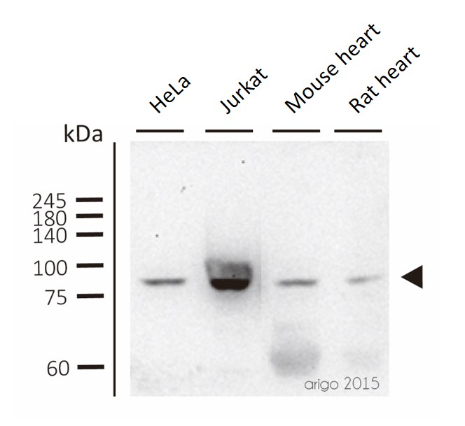
ARG55245 anti-WEE1 antibody WB image
Western blot: 30 μg of HeLa, Jurkat, Mouse heart and Rat heart lysates stained with ARG55245 anti-WEE1 antibody at 1:500 dilution.
-
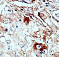
ARG55245 anti-WEE1 antibody IHC-P image
Immunohistochemistry: Formalin-fixed and paraffin-embedded Human cancer tissue stained with ARG55245 anti-WEE1 antibody.