ARG10740
anti-Visinin like 1 antibody
anti-Visinin like 1 antibody for ICC/IF,IHC-Frozen sections,Western blot and Human,Mouse,Rat,Bovine,Pig
Overview
| Product Description | Rabbit Polyclonal antibody recognizes Visinin like 1 |
|---|---|
| Tested Reactivity | Hu, Ms, Rat, Bov, Pig |
| Tested Application | ICC/IF, IHC-Fr, WB |
| Host | Rabbit |
| Clonality | Polyclonal |
| Isotype | IgG |
| Target Name | Visinin like 1 |
| Antigen Species | Human |
| Immunogen | Full length recombinant Human Visinin-like 1. |
| Conjugation | Un-conjugated |
| Alternate Names | HPCAL3; HUVISL1; Visinin-like protein 1; HLP3; Hippocalcin-like protein 3; VILIP; VLP-1; VILIP-1 |
Application Instructions
| Application Suggestion |
|
||||||||
|---|---|---|---|---|---|---|---|---|---|
| Application Note | * The dilutions indicate recommended starting dilutions and the optimal dilutions or concentrations should be determined by the scientist. |
Properties
| Form | Liquid |
|---|---|
| Purification | Affinity purification. |
| Buffer | PBS and 50% Glycerol. |
| Stabilizer | 50% Glycerol |
| Concentration | 1 mg/ml |
| Storage Instruction | For continuous use, store undiluted antibody at 2-8°C for up to a week. For long-term storage, aliquot and store at -20°C. Storage in frost free freezers is not recommended. Avoid repeated freeze/thaw cycles. Suggest spin the vial prior to opening. The antibody solution should be gently mixed before use. |
| Note | For laboratory research only, not for drug, diagnostic or other use. |
Bioinformation
| Database Links | |
|---|---|
| Gene Symbol | VSNL1 |
| Gene Full Name | visinin-like 1 |
| Background | This gene is a member of the visinin/recoverin subfamily of neuronal calcium sensor proteins. The encoded protein is strongly expressed in granule cells of the cerebellum where it associates with membranes in a calcium-dependent manner and modulates intracellular signaling pathways of the central nervous system by directly or indirectly regulating the activity of adenylyl cyclase. Alternatively spliced transcript variants have been observed, but their full-length nature has not been determined. [provided by RefSeq, Jul 2008] |
| Function | Regulates (in vitro) the inhibition of rhodopsin phosphorylation in a calcium-dependent manner. [UniProt] |
| Calculated MW | 22 kDa |
Images (4) Click the Picture to Zoom In
-
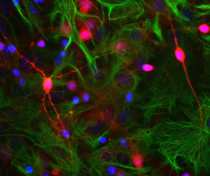
ARG10740 anti-Visinin like 1 antibody ICC/IF image
Immunocytochemistry: Rat Neuron-glia cell culture stained with ARG10740 anti-Visinin like 1 antibody (red) and co-stained with monoclonal antibody to GFAP (green); DNA (blue). ARG10740 reveals strong staining throughout some small neurons in soma and all processes, accumulation is seen in some hotspots along neurites. GFAP appears only in astrocytes.
-
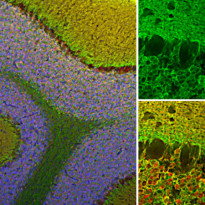
ARG10740 anti-Visinin like 1 antibody IHC-Fr image
Immunohistochemistry: Frozen section of Rat cerebellum stained with ARG10740 anti-Visinin like 1 antibody (green) at 1:2000 dilution and costained with ARG10703 anti-Calretinin antibody [6A9] (red) at 1:2000 dilution. DAPI (blue) for nuclear staining. (Sample preparation: Following transcardial perfusion of Rat with 4% paraformaldehyde, brain was post fixed for 24 hours, cut to 45 µM, and free-floating sections were stained with the above antibodies.)
The Visinin like 1 antibody reveals protein expressed in granule cells membranes and their synapses in both the granular and molecular layer of the cerebellum. The calretinin antibody stains the cytoplasm of neurons in the nuclear and molecular layers of cerebellum.
-
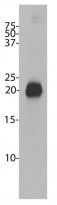
ARG10740 anti-Visinin like 1 antibody WB image
Western blot: Bovine cerebellum homogenate stained with ARG10740 anti-Visinin like 1 antibody.
-
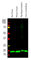
ARG10740 anti-Visinin like 1 antibody WB image
Western blot: Rat brain, Mouse brain, Pig hippocampus and Cow cerebellum lysates stained with ARG10740 anti-Visinin like 1 antibody (green) at 1:20000 dilution.


